. 2012 Nov 30;44(11):694-705.
doi: 10.3858/emm.2012.44.11.078.

IL-17-deficient allogeneic bone marrow transplantation prevents the induction of collagen-induced arthritis in DBA/1J mice

Affiliations

IL-17-deficient allogeneic bone marrow transplantation prevents the induction of collagen-induced arthritis in DBA/1J mice

Min Jung Park et al. Exp Mol Med. .

Abstract

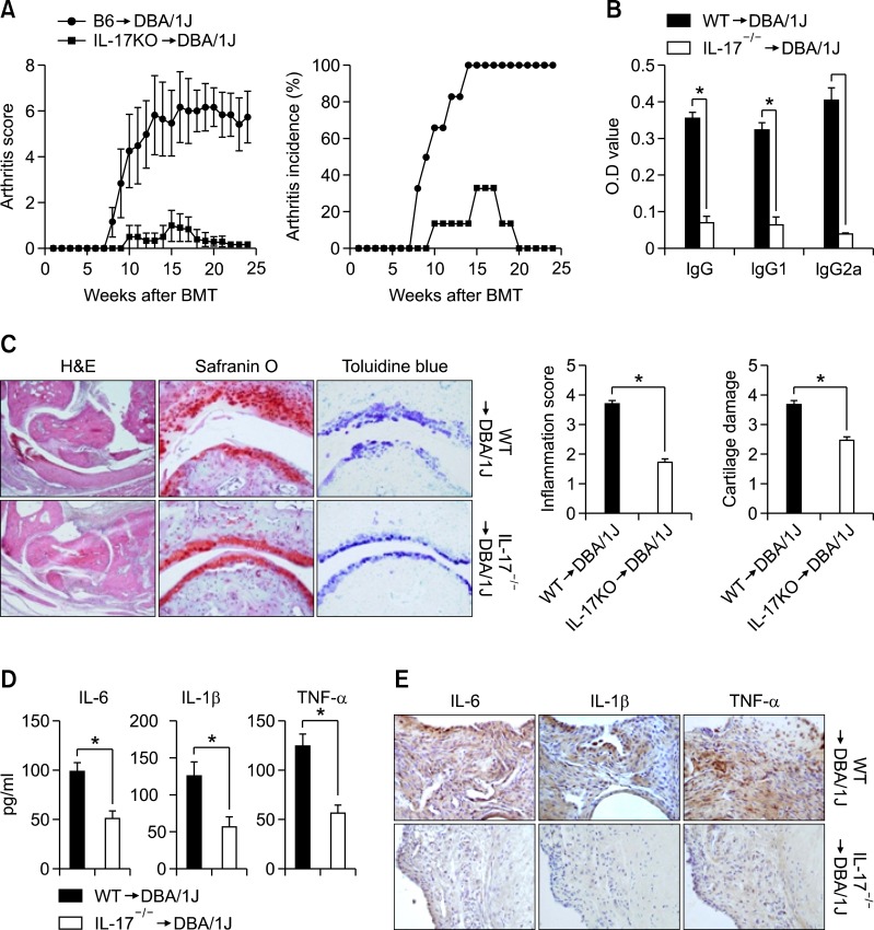
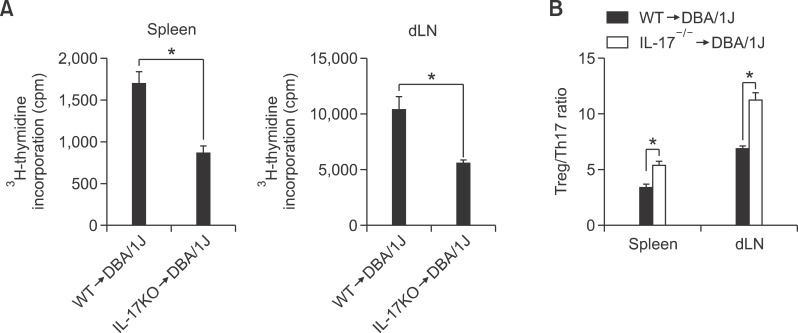
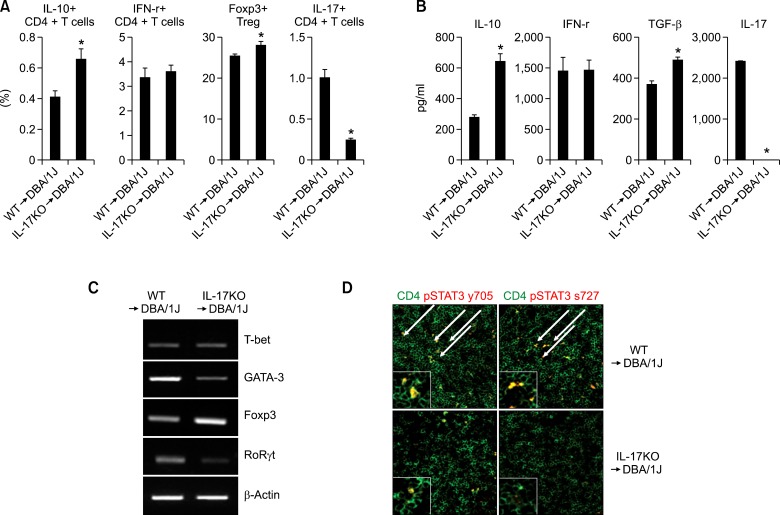
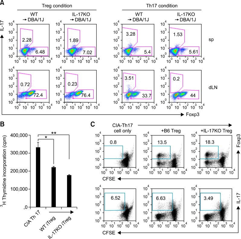
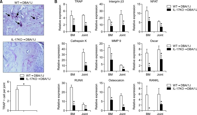
IL-17-producing CD4+ T cells (Th17) play important functions in autoimmune diseases and allograft rejection of solid organs. We examined the effects of IL 17 and its mechanism of action on arthritis in a murine collagen-induced arthritis (CIA) model using bone marrow transplantation (BMT) system. DBA/1J mice were administered a lethal radiation dose and then rescued with bone marrow derived from either wild-type (WT) or IL-17-/- mice on C57BL/6 background mice. CIA was induced after the bone marrow transplant, and disease progression was characterized. DBA/1J mice with CIA that received IL-17-/- donor bone marrow showed potently inhibited development and severity of clinical arthritis as compared with CIA mice that received WT bone marrow. Reduced secretion of the pro-inflammatory cytokines tumor necrosis factor-α, IL-1β, and IL-6, and collagen-specific T cell responses were observed in mice that received IL-17-/- bone marrow. IL-17 blockade also inhibited effector T cell proliferation by reciprocally regulating the Treg/Th17 ratio. IL-17 blockade prevented joint destruction in mice with CIA. These findings suggest that CIA with BMT is a viable method of immunological manipulation and that IL-17 deficiency suppresses severe joint destruction and inflammation in CIA mice. There may be clinical benefits in blocking IL-17 and BMT in the treatment of rheumatoid arthritis.


Figures

Figure 1
Figure 1
Reduced disease severity in IL-17 knockout (KO) → DBA chimeric mice. (A) Severity and incidence of arthritis in DBA/1J mice irradiated at 6 weeks and rescued with C57BL/6 (B6 → DBA) or interleukin (IL)-17-/- bone marrow (IL-17-/- → DBA). After the bone marrow transplant, mice were rested for 4 weeks and then collagen-induced arthritis (CIA) was initiated in transplant recipients (n = 10). Arthritis was monitored and scored as described in the Materials and Methods. Values are means ± SEM. (B) Levels of IgG, IgG1, and IgG2a antibodies to type II collagen (aCII) in serum from each group of mice 10 weeks after the primary immunization, as measured by ELISA. Values are means ± SEM of optical densities (OD). *P < 0.05. (C) Representative joint sections from each group of mice 8 weeks after the primary immunization. Left, hematoxylin and eosin (H&E) staining showing synovial inflammation. Original magnification, × 100. Middle, safranin O staining showing cartilage erosion. Original magnification, × 200. Right, toluidine blue staining. Original magnification, × 200. Graphs show the mean ± SEM inflammation and cartilage erosion scores for each group. *P < 0.05. (D) Inflammatory cytokine expression in bone marrow transplant (BMT) chimeric mice. The levels of cytokine in serum from each group of mice 10 weeks after the primary immunization, as measured by ELISA. Values are means ± SEM. *P < 0.05. (E) Joints obtained from each group examined for the expression of IL-1β, IL 6, and TNF-α by immunohistochemistry (× 200) or ELISA.
Figure 2
Figure 2
Type II collagen (CII) reactive T cell response in interleukin (IL)-17 knockout (KO)/DBA chimeric mice. (A) Proliferative response by spleen and draining lymph nodes (dLNs) from both chimeric mice, C57BL/6 (B6 → DBA) or IL-17-/- bone marrow (IL-17-/- → DBA) (n = 7 per group) 8 weeks after administration of the primary immunization. Spleen or dLN cells were cultured with type II collagen (CII) for 3 days. The proliferation response was measured by incorporation of 3H-thymidine. Graph shows the mean ± SEM. (B) Type II collagen (CII) specific Treg/Th17 regulation in spleen and draining lymph nodes (dLNs) from both chimeric mice. At 8 weeks after primary immunization, the spleens and dLN obtained from mice in each group were incubated with 40 g/ml of type II collagen (CII) for 3 days. CII-specific FoxP3:IL-17 ratio between CD4+ T cells in the spleen and dLN. Bars show the mean ± SEM. *P < 0.05 by Student's t-test.
Figure 3
Figure 3
Analysis of T helper cell population in interleukin (IL)-17 knockout (KO)/DBA chimeric mice. Cytokine production by CD4+ T cells from both chimeric mice, C57BL/6 (B6 → DBA) or IL-17-/- bone marrow (IL-17-/- → DBA) (n = 7 per group), 8 weeks after administration of the primary immunization. (A) Isolated CD4+ T cells were stimulated with anti-CD3 plus irradiated APC for 48 h. The percentages of IL-10, IFN-γ, Foxp3, and IL-17 expression in CD4+ T cells were determined via intracellular labeling plus flow cytometric analysis. (B) Production of cytokines in the culture medium was measured by ELISA. Data are shown as means ± SEM, n = 5-8 mice/group (*P < 0.05). Bars indicate combined means ± SD. *P < 0.05. (C) mRNA expression level of T cell transcription factors, T-bet (Th1), GATA-3 (Th2), Foxp3 (Treg), and RORgt (Th17) cells in the CD4+ T cells from both chimeric mice were analyzed by RT-PCR. (D) Confocal analysis of phospho-STAT3 expression in T cells. CD4 and pSTAT3 cells were stained with antibodies specific for CD4 (green) and phospho-STAT3 Tyr705 or Ser727 (red). Merged green and red is shown as yellow (pSTAT3 expression in CD4+ T cells). Original magnification, × 400 for panels D. All insets, ×1000.
Figure 4
Figure 4
Inhibition of effector T cell proliferation/response by interleukin (IL)-17 knockout (KO) bone marrow transplant (BMT)-induced Tregs. (A) CD4+ T cells were isolated from both chimeric mice, C57BL/6 (B6 → DBA) or IL-17-/- bone marrow (IL-17-/- → DBA), and cultured under activating conditions with Treg or Th17 polarizing conditions for 3 days, respectively. [Treg condition (Left): anti-CD3 and anti CD28 in the presence of recombinant hTGF-β, Th17 condition (Right): anti-CD28 in the presence of recombinant IL-6, TGF-β, anti-IL-4, and anti-IFN-γ]. Representative FACS plot depicting Foxp3 or IL-17 expression by CD4+ T cells. (B) CD4+ CD25+ T cells sorted from the above Treg condition cell cultures (a, left) were assessed for their suppressive capacity. CD4+ CD25+ T cells (5 × 10⁴ cells) were cultured with irradiated (5,000 rad) APCs (5 × 10⁴ cells) and Th17-conditioned culture T cells (5 × 10⁴ cells) isolated from CIA mice in the presence of CII for 72 h. The proliferation of effectors (Th17-conditioned T cells) was measured by incorporation of 3H-thymidine. Values are presented as means ± SEM. *P < 0.05, **P < 0.01. Data shown are representative of three independent experiments. (C) The regulatory effect of Treg in each group of mice. Treg were generated in vitro, sorted to > 99% purity, and cultured with effector cells. Treg cells from each group were cultured with CFSE-labeled CD4+ effector T cells from CIA mice. After 3 days, proliferation of effector cells co-cultured with Treg from B6 or IL-17KO was measured by CFSE dilution. Percentages of dividing effector cells (determined by CFSE incorporation) are shown. Data shown are representative of three independent experiments.
Figure 5
Figure 5
Inhibition of osteoclastogenesis in interleukin (IL)-17 knockout (KO)/DBA chimeric mice. (A) Tartrate-resistant acid phosphatase (TRAP) staining of paraffin sections from the joints of chimeric mice, C57BL/6 (B6 → DBA) or IL-17-/- bone marrow (IL-17-/- → DBA), showing multinucleated giant cells staining positive for TRAP. TRAP+ osteoclasts are indicated by black arrows. Original magnification × 200 in a. Bars show the mean ± SEM results of three experiments, each of which was performed in triplicate. *P < 0.01. (B) Real-time PCR analysis of osteoclast- and osteoblast-associated markers in joints and bone marrow. Relative gene expression was normalized against β-actin transcription levels. Each graph is representative of three independent experiments. *P < 0.01.

References

    1. Bettelli E, Oukka M, Kuchroo VK. T(H)-17 cells in the circle of immunity and autoimmunity. Nat Immunol. 2007;8:345–350. - PubMed
    1. Bluestone JA. Regulatory T-cell therapy: is it ready for the clinic? Nat Rev Immunol. 2005;5:343–349. - PubMed
    1. Camps M, Ruckle T, Ji H, Ardissone V, Rintelen F, Shaw J, Ferrandi C, Chabert C, Gillieron C, Francon B, Martin T, Gretener D, Perrin D, Leroy D, Vitte PA, Hirsch E, Wymann MP, Cirillo R, Schwarz MK, Rommel C. Blockade of PI3Kgamma suppresses joint inflammation and damage in mouse models of rheumatoid arthritis. Nat Med. 2005;11:936–943. - PubMed
    1. Chabaud M, Durand JM, Buchs N, Fossiez F, Page G, Frappart L, Miossec P. Human interleukin-17: A T cell-derived proinflammatory cytokine produced by the rheumatoid synovium. Arthritis Rheum. 1999;42:963–970. - PubMed
    1. Cho ML, Kang JW, Moon YM, Nam HJ, Jhun JY, Heo SB, Jin HT, Min SY, Ju JH, Park KS, Cho YG, Yoon CH, Park SH, Sung YC, Kim HY. STAT3 and NF-kappaB signal pathway is required for IL-23-mediated IL-17 production in spontaneous arthritis animal model IL-1 receptor antagonist-deficient mice. J Immunol. 2006a;176:5652–5661. - PubMed

Publication types

MeSH terms