Review
. 2012 Nov;122(11):3807-16.
doi: 10.1172/JCI58709. Epub 2012 Nov 1.

Lymphangioleiomyomatosis - a wolf in sheep's clothing

Affiliations
Review

Lymphangioleiomyomatosis - a wolf in sheep's clothing

Elizabeth P Henske et al. J Clin Invest. 2012 Nov.

Abstract

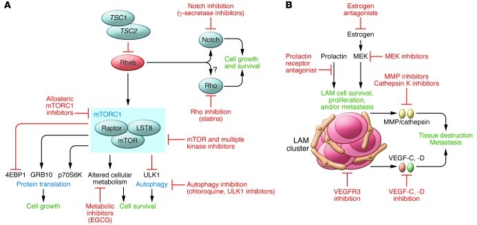
Lymphangioleiomyomatosis (LAM) is a rare progressive lung disease of women. LAM is caused by mutations in the tuberous sclerosis genes, resulting in activation of the mTOR complex 1 signaling network. Over the past 11 years, there has been remarkable progress in the understanding of LAM and rapid translation of this knowledge to an effective therapy. LAM pathogenic mechanisms mirror those of many forms of human cancer, including mutation, metabolic reprogramming, inappropriate growth and survival, metastasis via blood and lymphatic circulation, infiltration/invasion, sex steroid sensitivity, and local and remote tissue destruction. However, the smooth muscle cell that metastasizes, infiltrates, and destroys the lung in LAM arises from an unknown source and has an innocent histological appearance, with little evidence of proliferation. Thus, LAM is as an elegant, monogenic model of neoplasia, defying categorization as either benign or malignant.


Figures

Figure 1
Figure 1. The clinical and pathologic features of LAM.
(AD) Representative radiologic features of LAM include (A) multiple thin-walled cysts and pneumothorax, (B) pleural effusions (arrowhead), (C) renal angiomyolipomas (arrow), and (D) retroperitoneal lymphadenopathy (arrow). (E) High-power view of cystic change with surrounding LAM in the lung. LAM cells express the melanocystic antigen, HMB-45, as well as estrogen receptor and the smooth muscle cell antigen, smooth muscle actin. An immunohistochemical stain for podoplanin highlights lymphatic channels within cystic lesions and LAM cells clusters within the lymphatic lumen. Original magnification, ×400 (left and middle columns); ×200 (right column). Histology and immunohistochemistry courtesy of Kathryn Wikenheiser-Brokamp, Cincinnati Children’s Hospital Medical Center and the University of Cincinnati.
Figure 2
Figure 2. Signaling networks in LAM cells.
(A) LAM cells with biallelic mutational inactivation of the TSC1 or TSC2 gene have activation of the small GTPase and Ras homolog, Rheb. Rheb activates the “canonical” mTORC1 signaling network, leading to increased protein translation and decreased autophagy. Sirolimus (rapamycin) inhibits some of the functions of mTORC1. The impact of sirolimus on the targets of TORC1 may be cell type–specific and kinetically dynamic. New targets of the kinase domain of TORC1 continue to be identified. (B) Several TORC1-independent signaling functions of the TSC-Rheb node have been proposed, including activation of Notch and Rho and inhibition of B-Raf. Candidate TORC1-independent cellular activities of TSC-Rheb include aggresome accumulation and primary cilium formation.
Figure 3
Figure 3. Hypothesis for LAM progression.
LAM cells have smooth muscle cell features and originate from an unknown source; renal angiomyolipomas and uterine lesions have been proposed as potential primaries. These cells proliferate and drive a lymphangiogenic program that results in demarcation of tissue by chaotic lymphatic channels and the formation of LAM cell islands surrounded by lymphatic endothelium, which then bud into the lumen of the lymphatic system (i). These LAM cell clusters ascend the lymphatic tree by serial cycles of implantation and shedding (ii) and are transported by lymphatic flow to the venous circulation (iii) and ultimately impact in the pulmonary microvasculature (iv). Modified from ref. .
Figure 4
Figure 4. Mechanisms of airspace enlargement in LAM.
Two models of airspace enlargement in LAM are presented; these may not be mutually exclusive. (A) LAM cells secrete proteases including MMPs and cathepsin K, which degrade the extracellular matrix and induce apoptosis of alveolar epithelial cells. (B) LAM cells express lymphangiogenic growth factors, VEGF-C and VEGF-D, recruit lymphatic endothelial cells, drive the formation of lymphatic vascular channels and distort the lung architecture. Original magnification, ×200.
Figure 5
Figure 5. Future directions in therapy for LAM.
(A) Potential cell-autonomous therapeutic approaches in LAM include TORC1 inhibitors that may more effectively inhibit TORC1 (including kinase domain inhibitors) and/or have favorable toxicity and/or pharmacokinetic features; autophagy inhibitors; inhibitors of the putative “noncanonical” functions of TSC and Rheb, including Notch activation and Rho activation; direct inhibitors of Rheb’s activity (such as farnesyl transferase inhibitors). (B) Potential non-cell-autonomous therapeutic targets in LAM include inhibition of the lymphatic recruitment and vascular remodeling via inhibition of VEGF or VEGFR; inhibition of MMPs, cathepsin K, and other proteases that contribute to alveolar destruction; inhibition of LAM cells utilizing melanocyte or neural crest antigens as targets; and estrogen antagonism.

References

    1. Kitaichi M, Nishimura K, Itoh H, Izumi T. Pulmonary lymphangioleiomyomatosis: a report of 46 patients including a clinicopathologic study of prognostic factors. Am J Respir Crit Care Med. 1995;151(2 pt 1):527–533. - PubMed
    1. McCormack FX. Lymphangioleiomyomatosis: a clinical update. Chest. 2008;133(2):507–516. doi: 10.1378/chest.07-0898. - DOI - PubMed
    1. Johnson SR. Lymphangioleiomyomatosis. Eur Respir J. 2006;27(5):1056–1065. - PubMed
    1. Taveira-DaSilva AM, Steagall WK, Moss J. Lymphangioleiomyomatosis. Cancer Control. 2006;13(4):276–285. - PubMed
    1. Harknett EC, et al. Use of variability in national and regional data to estimate the prevalence of lymphangioleiomyomatosis. QJM. 2011;104(11):971–979. doi: 10.1093/qjmed/hcr116. - DOI - PubMed

Publication types

MeSH terms

Substances