. 2013 Feb;94(Pt 2):326-335.
doi: 10.1099/vir.0.046052-0. Epub 2012 Oct 31.

p53 controls hepatitis C virus non-structural protein 5A-mediated downregulation of GADD45α expression via the NF-κB and PI3K-Akt pathways

Affiliations

p53 controls hepatitis C virus non-structural protein 5A-mediated downregulation of GADD45α expression via the NF-κB and PI3K-Akt pathways

Du Cheng et al. J Gen Virol. 2013 Feb.

Abstract

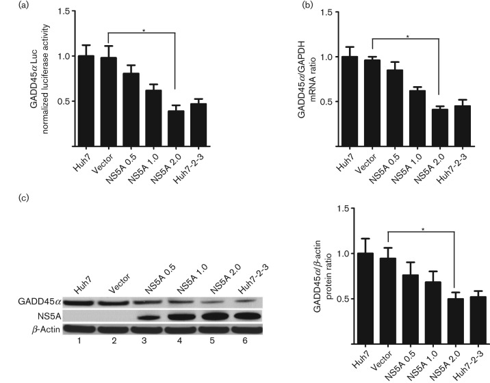
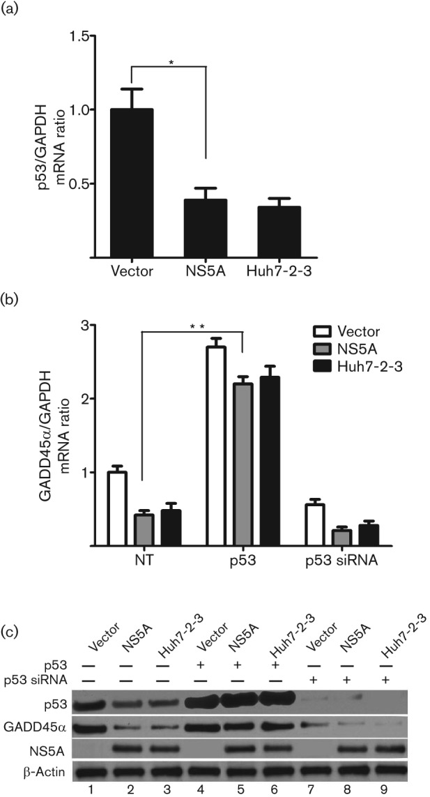
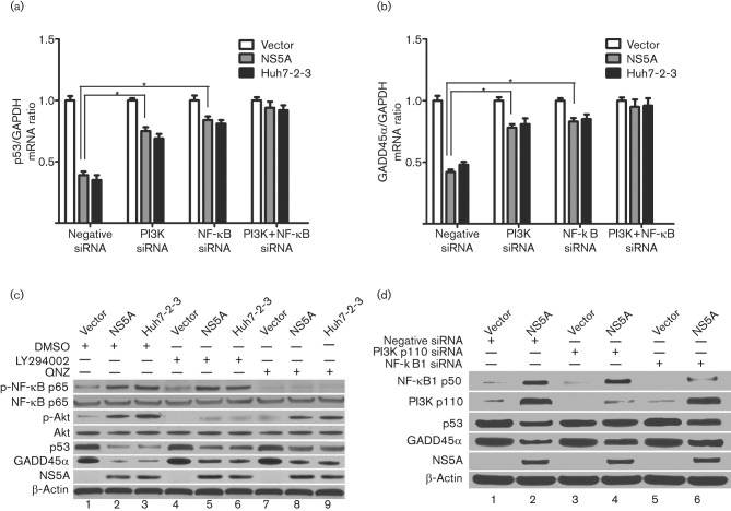
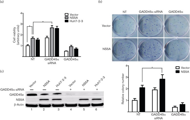
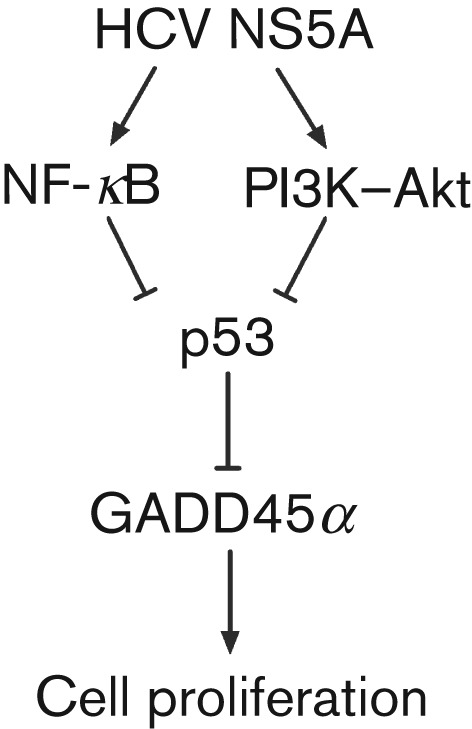
Growth arrest and DNA-damage-inducible gene 45-α (GADD45α) protein has been shown to be a tumour suppressor and is implicated in cell-cycle arrest and suppression of cell growth. The hepatitis C virus (HCV) non-structural 5A (NS5A) protein plays an important role in cell survival and is linked to the development of hepatocellular carcinoma (HCC). However, the role of HCV NS5A in the development of HCC remains to be clarified. This study sought to determine whether GADD45α mediates HCV NS5A-induced cellular survival and to elucidate the molecular mechanism of GADD45α expression regulated by HCV NS5A. It was found that HCV NS5A downregulated GADD45α expression at the transcriptional level by decreasing promoter activity, mRNA transcription and protein levels. Knockdown of p53 resulted in a similar decrease in GADD45α expression to that caused by HCV NS5A, whilst overexpression of p53 reversed the HCV NS5A-mediated downregulation of GADD45α. HCV NS5A repressed p53 expression, which was followed by a subsequent decrease in GADD45α expression. Further evidence was provided showing that HCV NS5A led to increases of phosphorylated nuclear factor-κB and Akt levels. Inhibition of these pathways using pharmacological inhibitors or specific small interfering RNAs rescued HCV NS5A-mediated downregulation of p53 and GADD45α. It was also found that HCV NS5A protein and depletion of GADD45α increased cell growth, whereas ectopic expression of GADD45α eliminated HCV NS5A-induced cell proliferation. These results indicated that HCV NS5A downregulates GADD45α expression and subsequently triggers cellular proliferation. These findings provide new insights suggesting that HCV NS5A could contribute to the occurrence of HCV-related HCC.


Figures

Fig. 1.
Fig. 1.
HCV NS5A downregulates GADD45α promoter activity, mRNA transcription and protein expression. (a) A GADD45α promoter–luciferase (Luc) reporter plasmid (1.0 µg) was co-transfected with the indicated plasmids at the indicated doses (µg) into Huh7 cells for 48 h and a luciferase assay was performed. (b) GADD45A mRNA levels were analysed by quantitative PCR. GADD45α mRNA levels were normalized to glyceraldehyde-3-phosphate dehydrogenase (GAPDH) levels to calculate the GADD45/GAPDH ratio, with the control non-transfected Huh7 cells having a ratio of 1. (c) Representative Western blot for GADD45α, NS5A and β-actin expression (left). Quantification of the GADD45α protein levels in Huh7-2-3 cells and in Huh7 cells transfected with NS5A-expressing vector or empty vector (right). Results are shown as means±sem of three independent experiments. *P<0.05.
Fig. 2.
Fig. 2.
p53 mediates HCV NS5A-induced downregulation of GADD45α expression. Stable vector-Huh7, NS5A-Huh7 and Huh7-2-3 cells were seeded into a 24-well plate 24 h before transfection with a p53 plasmid (0.5 µg) or p53 siRNA (50 nM) for 72 h. (a) Quantitative PCR for p53 mRNA levels. *P<0.05. (b) Quantitative PCR for GADD45α mRNA expression in the presence of p53 overexpression (p53) or knockdown (p53 siRNA). **P<0.001. Results for (a) and (b) are shown as means±sem of three independent experiments. (c) Representative Western blotting for p53, GADD45α, NS5A and β-actin protein levels. p53 protein was knocked down by p53 siRNAs (lanes 7–9). NS5A protein was detected in NS5A-Huh7 (lanes 2, 5 and 8) and Huh7-2-3 cells (lanes 3, 6 and 9).
Fig. 3.
Fig. 3.
The combination of inhibition of PI3K and NF-κB further rescues p53 and GADD45α expression. Stable vector-Huh7, NS5A-Huh7 and Huh7-2-3 cells were seeded into a 24-well plate with PI3K and NF-κB siRNAs (50 nM), as indicated. The cells were incubated for 72 h before harvest. (a, b) Effect of siRNAs targeting PI3K and NF-κB on p53 (a) and GADD45 (b) mRNA levels, determined by quantitative PCR. Results are shown as means±sem of three independent experiments. *P<0.05. (c) Effect of NF-κB activation inhibitor QNZ and PI3K inhibitor LY294002 on the phosphorylation of NF-κB and Akt and on the protein levels of p53 and GADD45α, determined by Western blotting. Cells were incubated with QNZ (5 µM) and LY294002 (50 µM) for 12 h before harvest. (d) Effect of siRNAs targeting PI3K and NF-κB1 on the protein levels of p53 and GADD45α determined by Western blotting. The expression levels of NF-κB1 and PI3K protein were knocked down by the respective siRNAs.
Fig. 4.
Fig. 4.
Role of GADD45α in HCV NS5A-induced cell proliferation. (a) Stable vector-Huh7, NS5A-Huh7 and Huh7-2-3 cells were seeded into a 96-well plate 24 h before transfection with GADD45α siRNA (50 nM) or GADD45α plasmid (0.5 µg), as indicated. The cells were incubated for 72 h before the cell viability assay. Results are shown as means±sem of three independent experiments. *P<0.05. (b) The effect of GADD45α on cellular growth was further confirmed by a colony formation assay. The upper panel shows representative images of colony formation in stable vector-Huh7 and NS5A-Huh7 cells transfected with GADD45α plasmid or GADD45α siRNAs. Quantitative analysis of colony numbers is shown in the lower panel (means±sem of three independent experiments). (c) Western blotting confirmed that the GADD45α protein was knocked down by GADD45α siRNAs. The NS5A protein was detected in stable NS5A-Huh7 and Huh7-2-3 cells.
Fig. 5.
Fig. 5.
Proposed mechanism for the downregulation of GADD45α expression by HCV NS5A, contributing to cell survival in human HCC cells. The NF-κB and PI3K–Akt pathways cooperatively influence HCV NS5A-mediated downregulation of p53 and subsequently inhibit GADD45α expression. The resulting GADD45α reduction contributes to cell proliferation.

References

    1. Boehme K. A., Kulikov R., Blattner C. (2008). p53 stabilization in response to DNA damage requires Akt/PKB and DNA-PK. Proc Natl Acad Sci U S A 105, 7785–7790 10.1073/pnas.0703423105 - DOI - PMC - PubMed
    1. Burdette D., Olivarez M., Waris G. (2010). Activation of transcription factor Nrf2 by hepatitis C virus induces the cell-survival pathway. J Gen Virol 91, 681–690 10.1099/vir.0.014340-0 - DOI - PubMed
    1. Cantley L. C. (2002). The phosphoinositide 3-kinase pathway. Science 296, 1655–1657 10.1126/science.296.5573.1655 - DOI - PubMed
    1. Chen S. L., Morgan T. R. (2006). The natural history of hepatitis C virus (HCV) infection. Int J Med Sci 3, 47–52 10.7150/ijms.3.47 - DOI - PMC - PubMed
    1. Chen F., Zhang Z., Leonard S. S., Shi X. (2001). Contrasting roles of NF-κB and JNK in arsenite-induced p53-independent expression of GADD45α. Oncogene 20, 3585–3589 10.1038/sj.onc.1204442 - DOI - PubMed

Publication types

MeSH terms

LinkOut - more resources