Review
. 2013 Jun;32(1-2):269-87.
doi: 10.1007/s10555-012-9413-5.

Heading off with the herd: how cancer cells might maneuver supernumerary centrosomes for directional migration

Affiliations
Review

Heading off with the herd: how cancer cells might maneuver supernumerary centrosomes for directional migration

Angela Ogden et al. Cancer Metastasis Rev. 2013 Jun.

Abstract

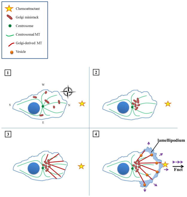
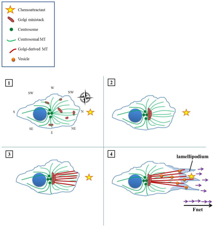
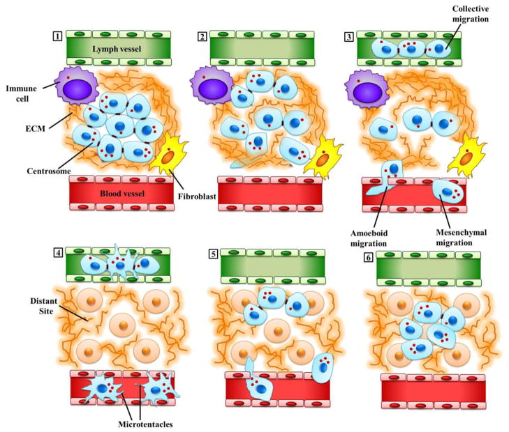
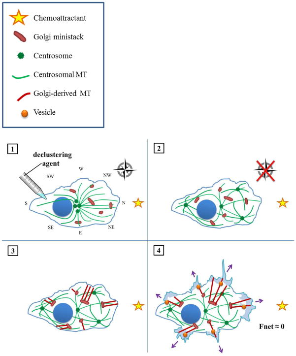
The complicity of centrosomes in carcinogenesis is unmistakable. Mounting evidence clearly implicates a robust correlation between centrosome amplification (CA) and malignant transformation in diverse tissue types. Furthermore, CA has been suggested as a marker of cancer aggressiveness, in particular the invasive phenotype, in breast and prostate cancers. One means by which CA promotes malignancy is through induction of transient spindle multipolarity during mitosis, which predisposes the cell to karyotypic changes arising from low-grade chromosome mis-segregation. It is well recognized that during cell migration in interphase, centrosome-mediated nucleation of a radial microtubule array is crucial for establishing a polarized Golgi apparatus, without which directionality is precluded. The question of how cancer cells maneuver their supernumerary centrosomes to achieve directionality during cell migration is virtually uncharted territory. Given that CA is a hallmark of cancers and has been correlated with cancer aggressiveness, malignant cells are presumably competent in managing their centrosome surfeit during directional migration, although the cellular logistics of this process remain unexplored. Another key angle worth pondering is whether an overabundance of centrosomes confers some advantage on cancer cells in terms of their migratory and invasive capabilities. Recent studies have uncovered a remarkable strategy that cancer cells employ to deal with the problem of excess centrosomes and ensure bipolar mitoses, viz., centrosome clustering. This review aims to change the narrative by exploring how an increased centrosome complement may, via aneuploidy-independent modulation of the microtubule cytoskeleton, enhance directional migration and invasion of malignant cells. We postulate that CA imbues cancer cells with cytoskeletal advantages that enhance cell polarization, Golgi-dependent vesicular trafficking, stromal invasion, and other aspects of metastatic progression. We also propose that centrosome declustering may represent a novel, cancer cell-specific antimetastatic strategy, as cancer cells may rely on centrosome clustering during migration as they do in mitosis. Elucidation of these details offers an exciting avenue for future research, as does investigating how CA may promote metastasis through enhanced directional migration.


Figures

Figure 1
Figure 1. Directional cell migration in the presence of a normal complement of centrosomes
(1) In an interphasic pre-migratory cell with a numerically and functionally normal centrosome complement, Golgi ministacks are scattered throughout the cytosol. The centrosome nucleates a cell-wide, radially-symmetric array of microtubules, which serves as a cellular compass (here, illustrated as the cardinal directions north, east, south, and west [49]). A chemoattractant (yellow star) is situated arbitrarily due north. The compass may persist throughout migration but is omitted from subsequent frames for reasons of clarity. (2) Golgi ministacks are collected into a distinct, polarized mass between the centrosome and leading edge, as directed by the centripetal signal provided by the radial microtubule array. (3) The Golgi serves as an MTOC and nucleates an asymmetric array of microtubules directed towards the leading edge of the cell (here, assigned northward for directional migration towards a hypothetical Northern stimulus). (4) The Golgi-nucleated microtubule array serves as tracks to the LE for the transport of vesicles laden with factors necessary for migration (such as focal adhesion- and actin-regulating proteins) as well as invasion (such as matrix metalloproteinases). Vesicles are not directed solely due North; there is also some lateral delivery owing to the coarseness of the compass. As a result, some of the forces driving cell migration (purple arrows) cancel out, but the net force vector (bold black arrow) still points northward, bringing the cell closer to its chemoattracting stimulus.
Figure 2
Figure 2. Enhanced directional cell migration in the presence of amplified centrosomes
(1) In the interphase, pre-migratory cell exhibiting CA, Golgi ministacks are likewise scattered throughout the cytosol. The supernumerary centrosomes nucleate a similarly cell-wide, radially-symmetric array of microtubules; however, microtubule density is increased owing to the increased number of MTOCs as well as enhanced microtubule-nucleating capacity of individual MTOCs. The resultant amplified microtubule array constitutes an enhanced cellular compass (illustrated here as having, for instance, additional SW, NW, SE, and NE ordinal directions). A chemoattractant is arbitrarily situated due North. (2) Golgi ministacks are collected into a more compact, polarized, continuous mass in between the centrosomes and leading edge, as the cell is better able to gauge the precise location of the stimulus. (3) The Golgi directs its microtubules more precisely towards the stimulus, resulting in a more focused array. (4) Vesicles with migration- and invasion-promoting factors travel along microtubule tracks to a more well-defined point due North, allowing more forces (purple arrows) to be directed towards the stimulus and finally sum to a greater net force (bold black arrow). Thus, the cell can accelerate towards its stimulus more expeditiously.
Figure 3
Figure 3. Extra centrosomes may promote directional migration and metastasis in malignant cells
(1) Malignant cells (depicted in light blue) are shown here in a cross-section through a primary tumor in situ. Some of these cells may have acquired extra centrosomes due to genetic mutations and other aberrant processes, while others, which may be distant from blood vessels and experience acute hypoxia, may have undergone hypoxia-induced CA. Tumor cells are connected to each other through cell-cell contacts, and are embedded within and interact extensively with the tumor microenvironment. Signals emanating from both the acellular components (including the ECM, depicted here as a brown fibrous matrix, and intratumoral conditions such as hypoxia) as well as the cellular components (such as fibroblasts, depicted here in yellow and immune cells, depicted in purple) of the tumor microenvironment, can potently influence various aspects of malignant cell behavior, including migration. (2) Initiation of cell migration from the primary tumor and invasion of the surrounding stroma involve the formation of actin polymerization-driven cellular protrusions. Supernumerary centrosomes may endow the cells bearing them with an enhanced radial signal to organize a compact, polarized Golgi apparatus which can efficiently direct vesicular traffic to the protrusions allowing more efficient delivery of enzymes for digesting the basement membrane, degrading ECM and carving paths for cell movement through the ECM. In addition, migration of such “super-centrosomal cells” may be enhanced by an “augmented” microtubule cytoskeleton characterized by increased microtubule density, more intense centrosome-dependent signaling and more robust nuclear-cytoskeletal connections. (3) Malignant cells exhibit great plasticity in the migratory modes they employ: they are capable of moving through interstitial spaces and intravasating into blood vessels (shown in red) or the lymphatic system (shown in green) either individually (amoeboid or mesenchymal) or collectively (the migration of cohesive multicellular units). ECM pore dimensions, ligand density, fibril stiffness and orientation together with cellular determinants such as cell-cell and cell-matrix adhesion, signals from tumor microenvironment and pericellular proteolysis, all influence the choice of migration mode. (4) The tumor cells circulate in the patient’s vasculature. Extra microtubules may help these cells to resist the mechanical stress of circulation. Circulating tumor cells protrude microtentacles, a process which may be facilitated by extra microtubules and/or centrosomes. Microtentacles enhance circulating tumor cell survival by facilitating their aggregation, and also allow them to grasp the vessel endothelium to ease and expedite extravasation. (5) Tumor cells exit the circulation (extravasate) and colonize distant niches. (6) Tumor cells produce a secondary tumor.
Figure 4
Figure 4. How declustering agents may hinder directional migration in cancer cells with amplified centrosomes
(1) A cell harboring supernumerary centrosomes is treated with a declustering agent. (2) Following scattering of centrosomes throughout the cell, the once highly organized, radial array of microtubules devolves into disarray. Hence, the cell literally loses its bearings. As a result, the cell lacks the centripetal signal necessary to organize the Golgi apparatus into a central, polarized mass. (3) Owing to their lack of polarization, the scattered Golgi ministacks nucleate microtubule arrays in various directions. (4) The cell directs trans-Golgi trafficking of vesicles to multiple cellular poles. Consequently, the exerts forces without respect to direction (purple arrows), which sum to a negligible net force (bold black arrow). The cell does not move appreciably towards its stimulus.

References

    1. D’Assoro AB, Lingle WL, Salisbury JL. Centrosome amplification and the development of cancer. Oncogene. 2002;21(40):6146–53. - PubMed
    1. Lingle WL, Salisbury JL. Altered centrosome structure is associated with abnormal mitoses in human breast tumors. Am J Pathol. 1999;155(6):1941–51. - PMC - PubMed
    1. Chan JY. A clinical overview of centrosome amplification in human cancers. Int J Biol Sci. 2011;7(8):1122–44. - PMC - PubMed
    1. Prigozhina NL, Waterman-Storer CM. Decreased polarity and increased random motility in PtK1 epithelial cells correlate with inhibition of endosomal recycling. J Cell Sci. 2006;119(Pt 17):3571–82. - PubMed
    1. Basto R, et al. Centrosome amplification can initiate tumorigenesis in flies. Cell. 2008;133(6):1032–42. - PMC - PubMed

Substances

LinkOut - more resources