. 2013 May;44(5):743-9.
doi: 10.1016/j.humpath.2012.07.018. Epub 2012 Oct 29.

Expression of hepatocyte epidermal growth factor receptor, FAS and glypican 3 in EpCAM-positive regenerative clusters of hepatocytes, cholangiocytes, and progenitor cells in human liver failure

Affiliations

Expression of hepatocyte epidermal growth factor receptor, FAS and glypican 3 in EpCAM-positive regenerative clusters of hepatocytes, cholangiocytes, and progenitor cells in human liver failure

Alex Hattoum et al. Hum Pathol. 2013 May.

Abstract

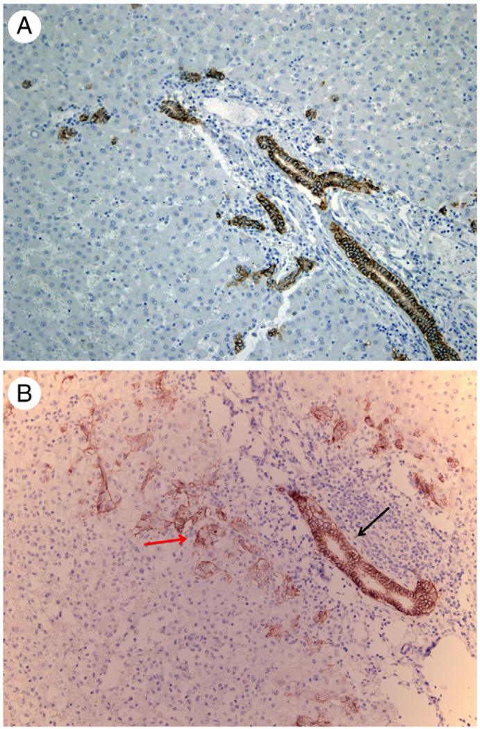
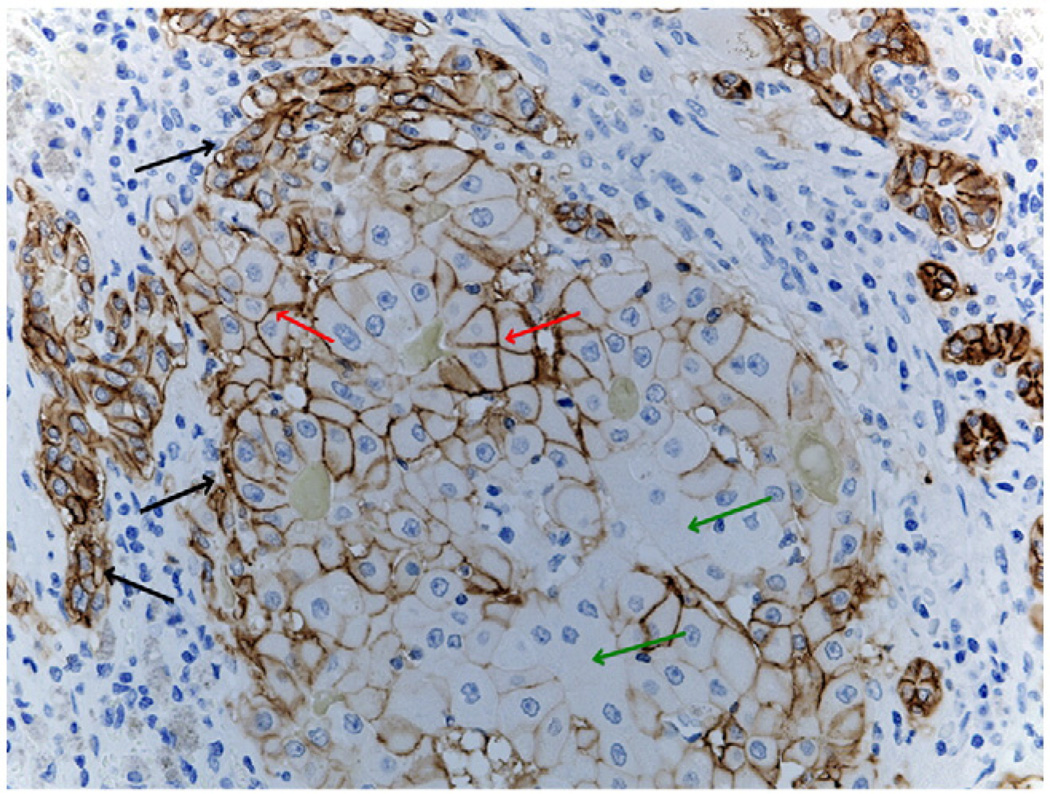
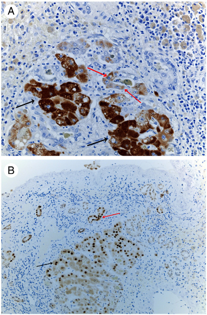
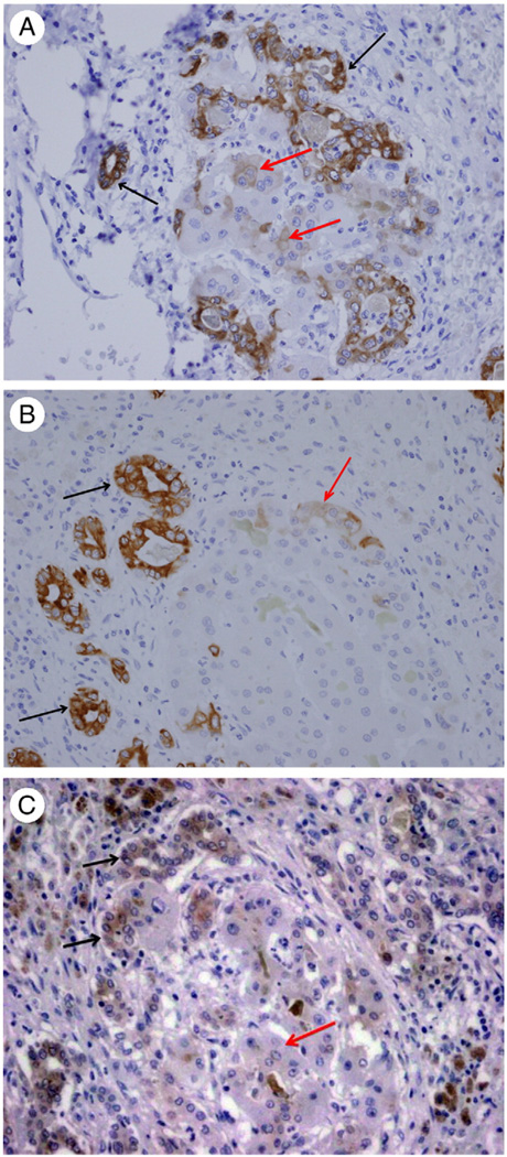
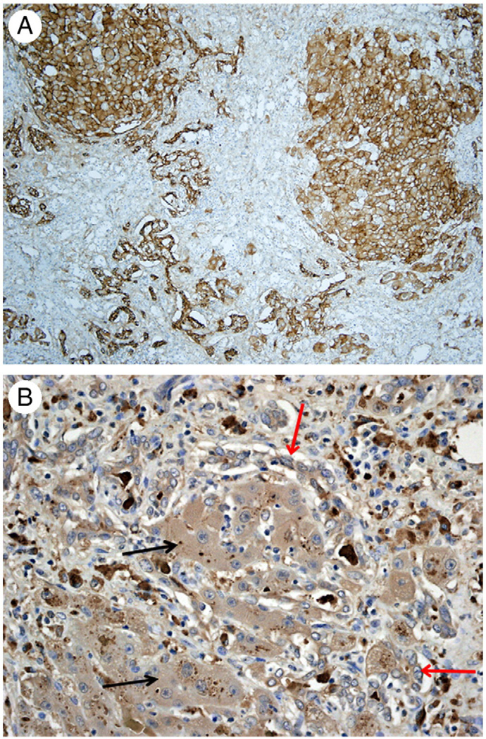
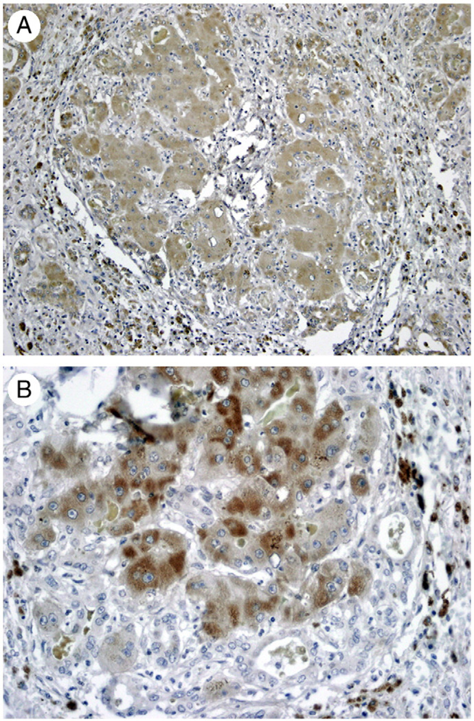
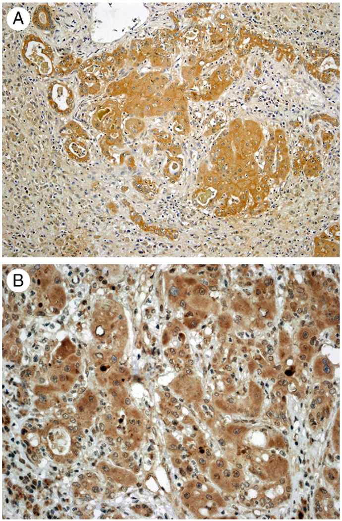
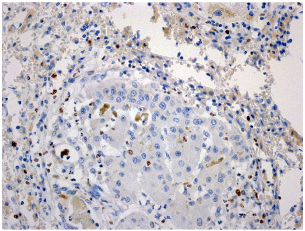
Liver regeneration under normal circumstances proceeds through proliferation of all cellular elements of the liver. Studies with rodent models have shown that when proliferation of hepatocytes is inhibited, progenitor cells arising from the biliary compartment transdifferentiate into "oval/progenitor" cells, which proceed to differentiate into hepatocytes. Recent studies have shown that the same pathways may operate in human liver failure. The growth factor receptors (HGF [hepatocyte growth factor] receptor) and epidermal growth factor receptor are key mitogenic receptors for both hepatocytes and progenitor cells. Our current study used the biliary and progenitor marker EpCAM (epithelial cell adhesion molecule) to detect "regenerative clusters" of mixed cholangiocyte-hepatocyte differentiation. We determined that expression of metabolic equivalent and epidermal growth factor receptor occurs in biliary cells, progenitor cells, and hepatocytes, whereas activation of metabolic equivalent and epidermal growth factor receptor is limited to regenerative cluster hepatocytes. These histologic events are associated with expression of apoptosis-inducing FAS and mitoinhibitory protein glypican 3. Cell proliferation was overall suppressed in regenerative clusters. Transdifferentiation of biliary and progenitor cells appears to be regulated by a complex interaction of signals promoting and arresting growth.


Figures

Fig. 1
Fig. 1
A, Immunohistochemistry for EpCAM on section from a normal human liver. Biliary ductules in portal triads and beyond (canals of Hering?) are the only cellular elements positive for EpCAM. Original magnification ×40. B, In a case of moderate hepatic tissue damage, a portal biliary ductule surrounded by inflammatory cells displays the standard strong positivity for EpCAM immunohistochemistry (black arrow). On either side of the portal space, numerous progenitor cells with histologic characteristics intermediate between biliary cells and hepatocytes also exhibit weaker EpCAM positivity (red arrow).
Fig. 2
Fig. 2
EpCAM immunohistochemistry in a regenerative cluster. Hepatobiliary cells in the periphery (black arrows) are strongly positive. Hepatocytes in the center display various degrees of immunoreactivity from strongly positive (red arrows) to completely negative (green arrows). (Original magnification ×100.)
Fig. 3
Fig. 3
A, Immunohistochemistry for hepatocyte-specific biomarker HEPPAR. Hepatocytes in the cluster stain positive (black arrows). HEPPAR-positive cells are also seen in cells embedded in ductules (red arrows). (Original magnification ×100.) B, Immunohistochemistry for hepatocyte-specific transcription factor HNF4α. Nuclei of cells recognizable as hepatocytes in the periphery of the cluster stain positive (black arrow). Positive nuclei are also seen, however, in biliary ductules in the periphery (red arrow). (Original magnification ×100.)
Fig. 4
Fig. 4
A, Immunohistochemistry for cytokeratin 7. Intense immunoreactivity is seen in cholangiocytes (black arrows). Some of the hepatocytes in the regenerative clusters also show weaker immunoreactivity (red arrows). (Original magnification ×100.) B, Immunohistochemistry for cytokeratin 19. Intense immunoreactivity is seen in cholangiocytes (black arrows). Some of the hepatocytes in the regenerative clusters also show weaker immunoreactivity (red arrow). (Original magnification ×100.) C, Immunoreactivity for NCAM1 (CD56) in a regenerative cluster. Positive immunoreactivity is confined to cholangiocytes in ductules (black arrows), whereas cells with hepatocyte morphology are negative (red arrows). (Original magnification ×100.)
Fig. 5
Fig. 5
A, Immunohistochemistry for EGFR. Both independent biliary ductules and adjacent regenerative clusters stain positive for EGFR. B, Immunohistochemistry for Tyr 1068–phosphorylated EGFR. Positive expression is seen in hepatocytes in the regenerative cluster (black arrows) compared with surrounding biliary ductules that are mostly negative (red arrows). (Original magnification ×100.)
Fig. 6
Fig. 6
A, Immunohistochemistry for HGF receptor MET. Both surrounding biliary ductules and hepatocytes of the regenerative cluster are positive. B, Immunohistochemistry for activated (Tyr 1003 phosphorylated) MET. Positive expression is seen in hepatocytes in the regenerative cluster, whereas surrounding biliary ductules are negative. (Original magnification ×100.)
Fig. 7
Fig. 7
Immunohistochemistry for death receptor FAS (A) and GPC3 (B). Both biliary cells and hepatocytes stain positive for these proteins. (Original magnification: A ×100, B ×200.)
Fig. 8
Fig. 8
Immunohistochemistry for Ki-67 in a regenerative cluster. Both hepatocytes and biliary cells are mostly negative for Ki-67. Occasional nonepithelial inflammatory cells stain positive (dark brown nuclei). (Original magnification ×100.)

References

    1. Limaye PB, Alarcon G, Walls AL, et al. Expression of specific hepatocyte and cholangiocyte transcription factors in human liver disease and embryonic development. Lab Invest. 2008;88:865–872. - PMC - PubMed
    1. Evarts RP, Nagy P, Nakatsukasa H, Marsden E, Thorgeirsson SS. In vivo differentiation of rat liver oval cells into hepatocytes. Cancer Res. 1989;49:1541–1547. - PubMed
    1. Nagy P, Bisgaard HC, Thorgeirsson SS. Expression of hepatic transcription factors during liver development and oval cell differentiation. J Cell Biol. 1994;126:223–233. - PMC - PubMed
    1. Golding M, Sarraf CE, Lalani EN, et al. Oval cell differentiation into hepatocytes in the acetylaminofluorene-treated regenerating rat liver. Hepatology. 1995;22:1243–1253. - PubMed
    1. Bisgaard HC, Nagy P, Santoni-Rugiu E, Thorgeirsson SS. Proliferation, apoptosis, and induction of hepatic transcription factors are characteristics of the early response of biliary epithelial (oval) cells to chemical carcinogens. Hepatology. 1996;23:62–70. - PubMed

Publication types

MeSH terms