

Overexpression of isocitrate dehydrogenase mutant proteins renders glioma cells more sensitive to radiation
- PMID: 23115158
- PMCID: PMC3534418
- DOI: 10.1093/neuonc/nos261
Overexpression of isocitrate dehydrogenase mutant proteins renders glioma cells more sensitive to radiation
Abstract
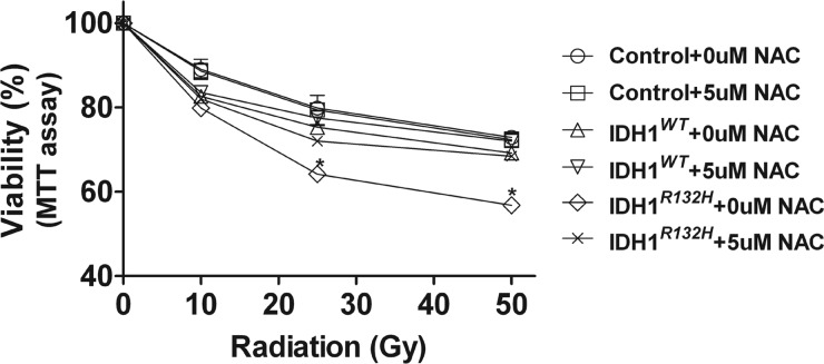
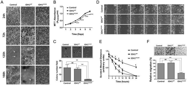
Mutations in isocitrate dehydrogenase 1 (IDH1) or 2 (IDH2) are found in a subset of gliomas. Among the many phenotypic differences between mutant and wild-type IDH1/2 gliomas, the most salient is that IDH1/2 mutant glioma patients demonstrate markedly improved survival compared with IDH1/2 wild-type glioma patients. To address the mechanism underlying the superior clinical outcome of IDH1/2 mutant glioma patients, we investigated whether overexpression of the IDH1(R132H) protein could affect response to therapy in the context of an isogenic glioma cell background. Stable clonal U87MG and U373MG cell lines overexpressing IDH1(WT) and IDH1(R132H) were generated, as well as U87MG cell lines overexpressing IDH2(WT) and IDH2(R172K). In vitro experiments were conducted to characterize baseline growth and migration and response to radiation and temozolomide. In addition, reactive oxygen species (ROS) levels were measured under various conditions. U87MG-IDH1(R132H) cells, U373MG-IDH1(R132H) cells, and U87MG-IDH2(R172K) cells demonstrated increased sensitivity to radiation but not to temozolomide. Radiosensitization of U87MG-IDH1(R132H) cells was accompanied by increased apoptosis and accentuated ROS generation, and this effect was abrogated by the presence of the ROS scavenger N-acetyl-cysteine. Interestingly, U87MG-IDH1(R132H) cells also displayed decreased growth at higher cell density and in soft agar, as well as decreased migration. Overexpression of IDH1(R132H) and IDH2(R172K) mutant protein in glioblastoma cells resulted in increased radiation sensitivity and altered ROS metabolism and suppression of growth and migration in vitro. These findings provide insight into possible mechanisms contributing to the improved outcomes observed in patients with IDH1/2 mutant gliomas.
Figures
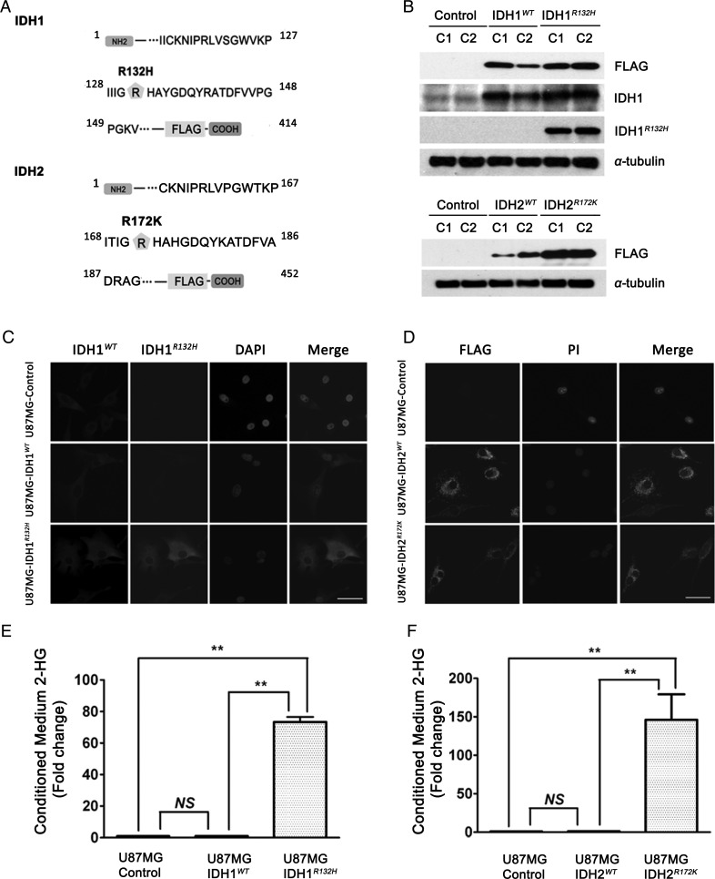
 ). (B) Representative western blot for FLAG-tagged IDH1WT and IDH1R132H or IDH2WT and IDH2R172K in stably transfected U87MG cells (2 representative independent clones for each construct, C1 and C2, are shown) using anti-FLAG, anti- IDH1WT, and anti-IDH1R132H antibodies. (C) Immunofluorescence showed the diffuse cytoplasmic distribution of IDH1WT and IDH1R132H proteins stained by anti-FLAG antibody (green), anti-IDH1WT antibody (red), and anti-IDH1R132H (green). Nuclei were counterstained with DAPI (blue). 40×, bar = 20μm (D) Immunofluorescence shows the punctate cytoplasmic distribution of IDH2WT and IDH2R172K proteins stained by anti-FLAG antibody (green), and nuclei were counterstained with propidium iodide (PI) (red), 40×, bar = 20μm. (E) ∼70-fold increase in 2-HG level was found in the conditioned medium of U87MG-IDH1R132H cells by HPLC-MS. Data were normalized by cell number and expressed as fold change relative to U87MG-control (mean ± SEM, n =3). **P < .01 compared with U87MG-control and U87MG-IDH1WT cells. (F) ∼140-fold increase in 2-HG level was found in the conditioned medium of U87MG-IDH2R172K cells by HPLC-MS. Data were normalized by cell number and expressed as mean fold change relative to U87MG-control (mean ± SEM, n =3). **P < .01 compared with U87MG-control and U87MG-IDH2WT cells.

References
-
- Balss J, Meyer J, Mueller W, Korshunov A, Hartmann C, von Deimling A. Analysis of the IDH1 codon 132 mutation in brain tumors. Acta Neuropathol. 2008;116(6):597–602. - PubMed
Publication types
MeSH terms
Substances
Grants and funding
LinkOut - more resources
Full Text Sources
Medical
Miscellaneous
