

Hippocampal perfusion predicts impending neurodegeneration in REM sleep behavior disorder
- PMID: 23115214
- PMCID: PMC3578380
- DOI: 10.1212/WNL.0b013e318278b658
Hippocampal perfusion predicts impending neurodegeneration in REM sleep behavior disorder
Abstract
Objectives: Patients with idiopathic REM sleep behavior disorder (IRBD) are at risk for developing Parkinson disease (PD) and dementia with Lewy bodies (DLB). We aimed to identify functional brain imaging patterns predicting the emergence of PD and DLB in patients with IRBD, using SPECT with (99m)Tc-ethylene cysteinate dimer (ECD).
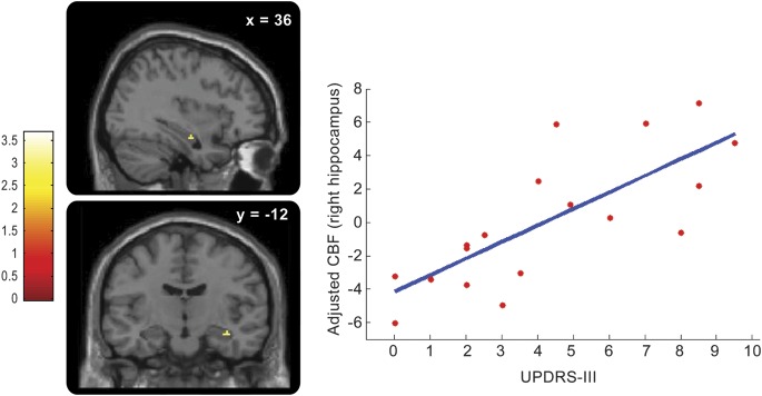
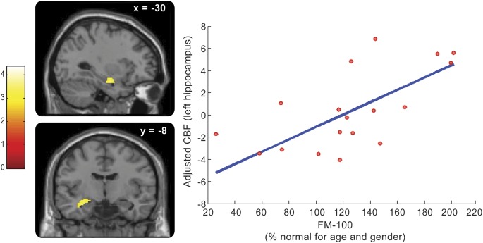
Methods: Twenty patients with IRBD were scanned at baseline during wakefulness using (99m)Tc-ECD SPECT. After a follow-up of 3 years on average, patients were divided into 2 groups according to whether or not they developed defined neurodegenerative disease (PD, DLB). SPECT data analysis comparing regional cerebral blood flow (rCBF) between groups assessed whether specific brain perfusion patterns were associated with subsequent clinical evolution. Regression analysis between rCBF and clinical markers of neurodegeneration (motor, color vision, olfaction) looked for neural structures involved in this process.
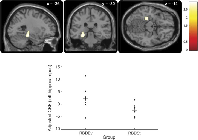
Results: Of the 20 patients with IRBD recruited for this study, 10 converted to PD or DLB during the follow-up. rCBF at baseline was increased in the hippocampus of patients who would later convert compared with those who would not (p < 0.05 corrected). Hippocampal perfusion was correlated with motor and color vision scores across all IRBD patients.
Conclusions: (99m)Tc-ECD SPECT identifies patients with IRBD at risk for conversion to other neurodegenerative disorders such as PD or DLB; disease progression in IRBD is predicted by abnormal perfusion in the hippocampus at baseline. Perfusion within this structure is correlated with clinical markers of neurodegeneration, further suggesting its involvement in the development of presumed synucleinopathies.
Figures
Comment in
-
Imaging markers offer promise: dream enactment may predict your patient's future.Neurology. 2012 Dec 11;79(24):2296-7. doi: 10.1212/WNL.0b013e318278b6b7. Epub 2012 Oct 31. Neurology. 2012. PMID: 23115206 No abstract available.
References
-
- Iranzo A, Molinuevo JL, Santamaria J, et al. Rapid-eye-movement sleep behaviour disorder as an early marker for a neurodegenerative disorder: a descriptive study. Lancet Neurol 2006;5:572–577 - PubMed
-
- Postuma RB, Gagnon JF, Vendette M, Desjardins C, Montplaisir JY. Olfaction and color vision identify impending neurodegeneration in rapid eye movement sleep behavior disorder. Ann Neurol 2011;69:811–818 - PubMed
-
- Iranzo A, Lomena F, Stockner H, et al. Decreased striatal dopamine transporter uptake and substantia nigra hyperechogenicity as risk markers of synucleinopathy in patients with idiopathic rapid-eye-movement sleep behaviour disorder: a prospective study. Lancet Neurol 2010;9:1070–1077 - PubMed
-
- Vendette M, Gagnon JF, Soucy JP, et al. Brain perfusion and markers of neurodegeneration in rapid eye movement sleep behavior disorder. Mov Disord 2011;26:1717–1724 - PubMed
-
- Cistaro A, Valentini MC, Chio A, et al. Brain hypermetabolism in amyotrophic lateral sclerosis: a FDG PET study in ALS of spinal and bulbar onset. Eur J Nucl Med Mol Imaging 2012;39:251–259 - PubMed
Publication types
MeSH terms
Grants and funding
LinkOut - more resources
Full Text Sources
Other Literature Sources