. 2012 Dec 21;287(52):43639-50.
doi: 10.1074/jbc.M112.393678. Epub 2012 Oct 31.

The mRNA-stabilizing factor HuR protein is targeted by β-TrCP protein for degradation in response to glycolysis inhibition

Affiliations

The mRNA-stabilizing factor HuR protein is targeted by β-TrCP protein for degradation in response to glycolysis inhibition

Po-Chen Chu et al. J Biol Chem. .

Abstract

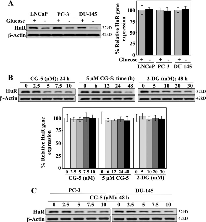
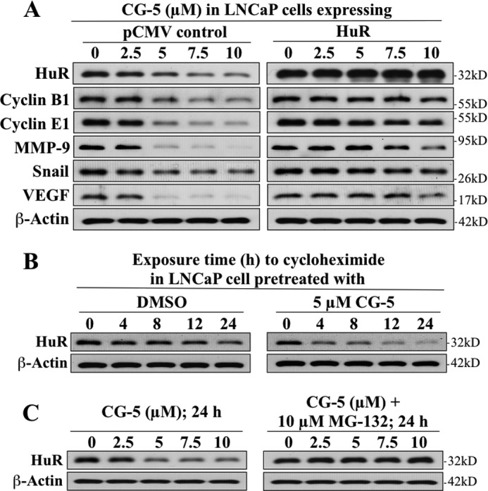
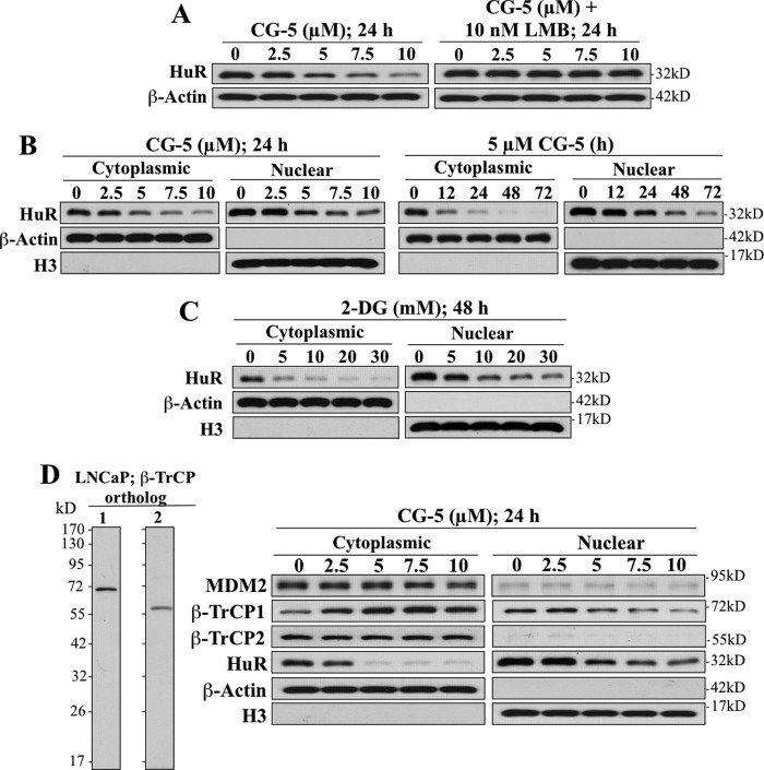
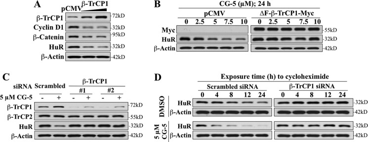
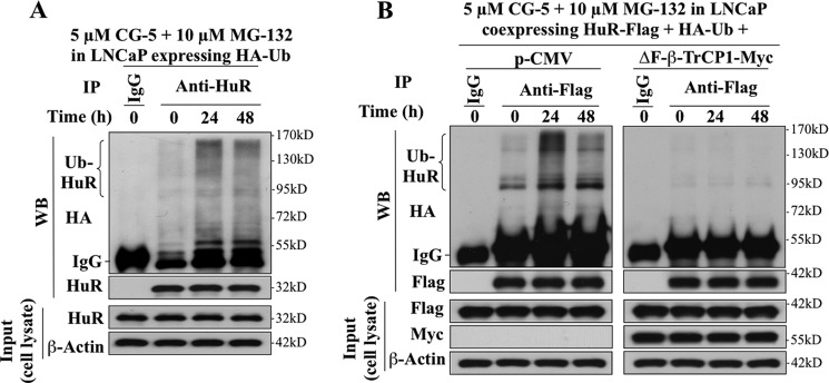
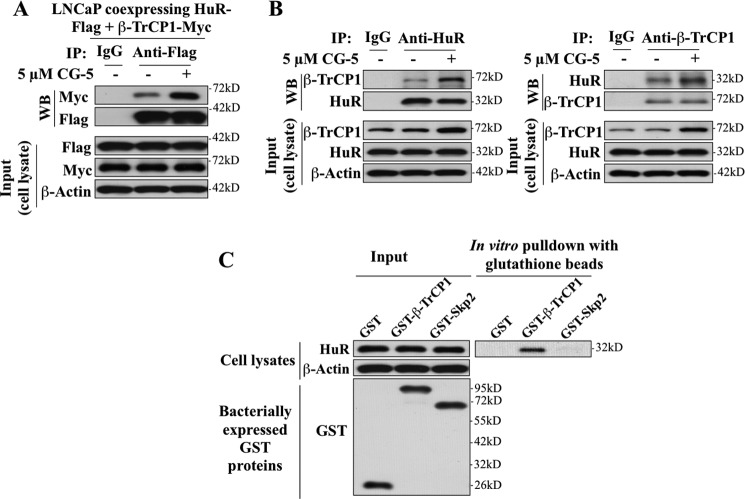
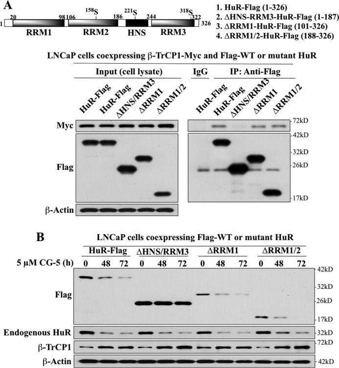
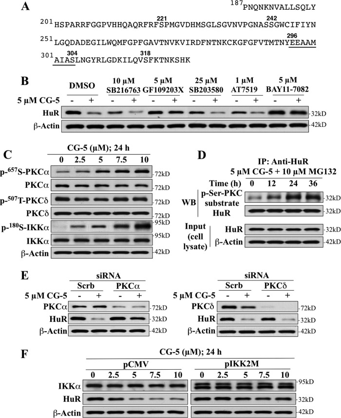
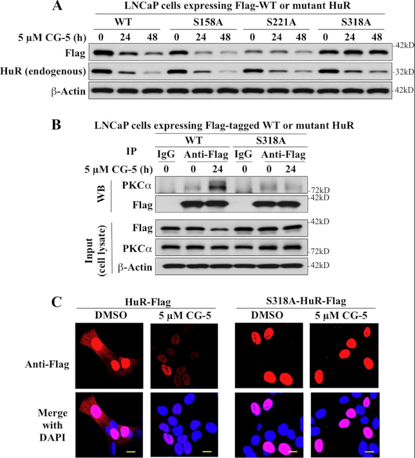
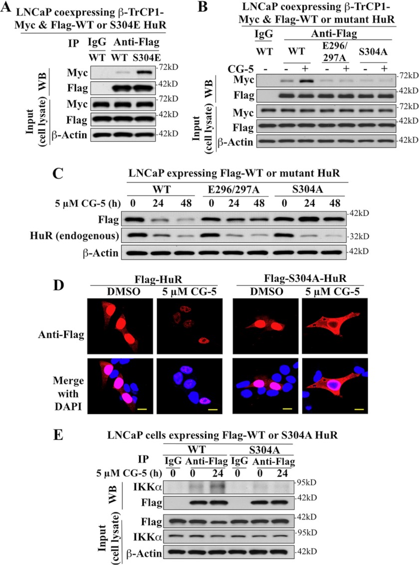
The mRNA-stabilizing protein HuR acts a stress response protein whose function and/or protein stability are modulated by diverse stress stimuli through posttranslational modifications. Here, we report a novel mechanism by which metabolic stress facilitates proteasomal degradation of HuR in cancer cells. In response to the glucose transporter inhibitor CG-5, HuR translocates to the cytoplasm, where it is targeted by the ubiquitin E3 ligase β-TrCP1 for degradation. The cytoplasmic localization of HuR is facilitated by PKCα-mediated phosphorylation at Ser-318 as the Ser-318 → alanine substitution abolishes the ability of the resulting HuR to bind PKCα and to undergo nuclear export. The mechanistic link between β-TrCP1 and HuR degradation was supported by the ability of ectopically expressed β-TrCP1 to mimic CG-5 to promote HuR degradation and by the protective effect of dominant negative inhibition of β-TrCP1 on HuR ubiquitination and degradation. Substrate targeting of HuR by β-TrCP1 was further verified by coimmunoprecipitation and in vitro GST pull-down assays and by the identification of a β-TrCP1 recognition site. Although HuR does not contain a DSG destruction motif, we obtained evidence that β-TrCP1 recognizes an unconventional motif, (296)EEAMAIAS(304), in the RNA recognition motif 3. Furthermore, mutational analysis indicates that IKKα-dependent phosphorylation at Ser-304 is crucial to the binding of HuR to β-TrCP1. Mechanistically, this HuR degradation pathway differs from that reported for heat shock and hypoxia, which underlies the complexity in the regulation of HuR turnover under different stress stimuli. The ability of glycolysis inhibitors to target the expression of oncogenic proteins through HuR degradation might foster novel strategies for cancer therapy.


Figures

FIGURE 1.
FIGURE 1.
Glycolysis inhibition suppresses HuR expression at the protein level in prostate cancer cells. A, Western blot (left panel) and qRT-PCR (right panel) analyses of the effect of glucose deprivation on the protein and mRNA expression levels of HuR in LNCaP, PC-3, and DU-145 cells in 10% FBS-supplemented, glucose-free RPMI 1640 medium for 72 h. B, Western blot (upper panel) and qRT-PCR (lower panel) analyses of dose- and/or time-dependent effects of CG-5 and 2-DG on the protein and mRNA expression levels of HuR in LNCaP cells. C, dose-dependent effects of CG-5 on HuR protein expression in PC-3 and DU-145 cells after 48 h of exposure.
FIGURE 2.
FIGURE 2.
Effects of CG-5 on the expression of HuR-targeted signaling proteins and HuR protein stability. A, validation of the role of HuR in mediating the suppressive effect of CG-5 on various tumor-promoting signaling proteins encoded by HuR-targeted mRNAs, including cyclin B1, cyclin E1, MMP9, Snail, and VEGF in LNCaP cells. Ectopic expression of full-length HuR protected cells from CG-5-mediated suppression of these proteins. B, cycloheximide chase assays of the effect of CG-5 on the half-life of HuR protein in LNCaP cells. Cells were treated with 5 μm CG-5 for 12 h, followed by exposure to 100 μg/ml cycloheximide for the indicated time intervals. C, protective effect of proteasome inhibitor MG132 on CG-5-mediated HuR degradation. LNCaP cells were treated with CG-5 at the indicated concentration, alone or in combination with 10 μm MG132, for 24 h.
FIGURE 3.
FIGURE 3.
CG-5-induced HuR degradation is preceded by nuclear export in LNCaP cells. A, the nuclear export inhibitor, leptomycin B (LMB), protected cells from CG-5-mediated HuR degradation. B, dose- and time-dependent suppressive effects of CG-5 on HuR expression in the cytoplasm versus nucleus. β-Actin and histone H3 signals indicate the quality of the cytoplasmic and nuclear fractionations, respectively. C, dose-dependent effect of 2-DG on the cytoplasmic and nuclear expression of HuR protein after 48 h of treatment. D, left panel, Western blot analysis of the expression of the two paralogs of β-TrCP, β-TrCP1 and β-TrCP2, in LNCaP cells. Right panel, dose-dependent effects of CG-5 on cytoplasmic and nuclear expression levels of putative HuR-targeted E3 ligases, including β-TrCP1, β-TrCP2, and MDM2.
FIGURE 4.
FIGURE 4.
Evidence that β-TrCP1 is involved in CG-5-mediated HuR degradation in LNCaP cells. A, effect of ectopically expressed full-length β-TrCP1 versus pCMV vector control on the expression of HuR and known β-TrCP substrates, cyclin D1 and β-catenin. B, protective effect of ΔF-β-TrCP1-Myc, a dominant-negative mutant form of β-TrCP1, against CG-5-mediated HuR degradation. C, protective effects of siRNA-mediated knockdown of β-TrCP1 by using two independent siRNAs (#1 and #2) on CG-5-mediated HuR degradation. D, cycloheximide chase analysis of the effect of siRNA-mediated knockdown of β-TrCP1 on HuR protein stability in response to DMSO versus 5 μm CG-5 LNCaP cells. Cells were electroporated with scrambled or β-TrCP1 siRNA. After incubation for 48 h, these cells were treated with 5 μm CG-5 for 12h, followed by exposure to 100 μg/ml cycloheximide for the indicated time intervals.
FIGURE 5.
FIGURE 5.
CG-5 facilitated β-TrCP1-mediated HuR ubiquitination in LNCaP cells. A, coimmunoprecipitation analysis of the time-dependent effect of CG-5 (5 μm) on HuR ubiquitination. LNCaP cells ectopically expressing HA-Ubiquitin (HA-Ub) were treated with 5 μm CG-5 for 12 or 36 h, followed by cotreatment with proteasome inhibitor MG132 for an additional 12 h. Equal amounts of cell lysates were immunoprecipitated (IP) with anti-HuR antibody and protein A/G-agarose followed by immunoblotting (WB) with anti-HA antibodies. B, ectopic expression of ΔF-β-TrCP1-Myc blocked CG-5-mediated HuR ubiquitination. LNCaP cells cotransfected with plasmids encoding HA-Ub, HuR-FLAG, and ΔF-β-TrCP1-Myc or empty vector (pCMV) were treated with 5 μm CG-5 for 12 or 36 h, followed by cotreatment with proteasome inhibitor MG132 for an additional 12 h. Equal amounts of cell lysates were immunoprecipitated with anti-FLAG antibody and protein A/G-agarose followed by immunoblotting with anti-HA and anti-FLAG antibodies.
FIGURE 6.
FIGURE 6.
Evidence that HuR physically interacts with β-TrCP1. A, coimmunoprecipitation analysis revealed the association of HuR with β-TrCP1 in response to CG-5 treatment. LNCaP cells ectopically expressing both FLAG-tagged HuR and Myc-tagged β-TrCP1 were treated with 5 μm CG-5 for 12 h, followed by cotreatment with 10 μm MG132 for an additional 12 h. Equal amounts of cell lysates were immunoprecipitated (IP) with anti-FLAG antibody and protein A/G-agarose followed by immunoblotting (WB) with anti-Myc and anti-FLAG antibodies. B, coimmunoprecipitation analysis showed the association of endogenous HuR and β-TrCP1 in response to CG-5 treatment. Cells were treated according to the aforementioned procedure. Equal amounts of cell lysates were immunoprecipitated with anti-HuR (left panel) and anti-β-TrCP1 (right panel) antibodies and protein A/G-agarose, followed by immunoblotting with anti-β-TrCP1 and anti-HuR antibodies, respectively. C, in vitro pull-down of HuR by bacterially expressed GST-β-TrCP1. Equal amounts of LNCaP cell lysates were incubated with recombinant GST, GST-β-TrCP1, or GST-Skp2 immobilized onto glutathione beads. The resulting complexes were washed, centrifuged, and subjected to immunoblotting analysis with HuR antibody (right panel). One-tenth volume of cell lysates were collected as input and probed with HuR and β-actin antibodies, and recombinant GST-fusion proteins were purified and probed with GST antibody (left panel).
FIGURE 7.
FIGURE 7.
Evidence that the HNS-RRM3 motif is involved in the recognition and degradation of HuR by β-TrCP1. A, upper panel, schematic representation of the structures of wild-type and various truncated mutant forms of HuR-FLAG. Lower panel, coimmunoprecipitation analysis of the interaction of wild-type HuR versus various truncated HuR mutants with β-TrCP1. LNCaP cells were transiently cotransfected with Myc-tagged β-TrCP1 and FLAG-tagged wild-type HuR or individual truncated mutants for 48 h. Immunoprecipitation (IP) with anti-FLAG-agarose conjugates and immunoblotting with anti-Myc and anti-FLAG antibodies were performed as described in Fig. 6. B, time-dependent effect of CG-5 on the degradation of FLAG-tagged HuR and various truncated HuR mutants versus endogenous HuR.
FIGURE 8.
FIGURE 8.
Evidence that PKCα and IKKα play pivotal roles in CG-5-facilitated HuR degradation in LNCaP cells. A, the amino acid sequence of the HNS-RRM3 motif of HuR. B, effects of various kinase inhibitors, including SB216763 (GSK3β), GF109203X (PKC), SB203580 (p38), AT7519 (CDK), and BAY11–7082 (IKKα) on CG-5-mediated HuR degradation. Cells were treated with CG-5 in combination with individual kinase inhibitors at the indicated concentrations for 24 h. C, dose-dependent effect of CG-5 on the phosphorylation status of PKCα, PKCδ, and IKKα in LNCaP cells. D, coimmunoprecipitation analysis of the time-dependent effect of 5 μm CG-5 on PKC-mediated serine phosphorylation of HuR. Cells were treated with 5 μm CG-5 for 12 or 24 h followed by cotreatment with 10 μm MG132 for an additional 12 h. Equal amounts of cell lysates were immunoprecipitated (IP) with anti-HuR antibody and protein A/G-agarose followed by immunoblotting (WB) with anti-p-Ser PKC substrate and anti-HuR antibodies. E, effects of siRNA-mediated silencing of PKCα versus PKCδ on CG-5-mediated HuR degradation. Cells were transfected with 100 nm scrambled (Scrb), PKCα (left panel), or PKCδ (right panel) siRNA for 48 h and were then treated with 5 μm CG-5 for 24 h. F, dominant-negative inhibition of IKKα by IKK2M protected HuR from CG-5-mediated degradation.
FIGURE 9.
FIGURE 9.
Mutational analyses demonstrating the crucial role of Ser-318 in CG-5-mediated HuR cytoplasmic translocation and degradation in LNCaP cells. A, effect of alanine substitution at Ser-158, Ser-221, and Ser-318 on CG-5-mediated HuR degradation. B, coimmunoprecipitation analysis of the effect of the Ser-318 → Ala substitution on HuR binding to PKCα. LNCaP cells ectopically expressing FLAG-tagged WT or S318A mutant form of HuR were treated with 5 μm CG-5 for 24 h. Immunoprecipitation (IP) with anti-FLAG-agarose conjugates and immunoblotting (WB) with anti- PKCα and anti-FLAG antibodies were performed as described in Fig. 6. C, immunocytochemical analysis of the cellular distribution and turnover of FLAG-tagged WT versus the S318A mutant of HuR in CG-5-treated cells. Scale bars = 10 μm.
FIGURE 10.
FIGURE 10.
Identification of the β-TrCP1 recognition motif in HuR in LNCaP cells via mutational analyses. A, phosphomimetic substitution of Ser-304 by Glu promotes the binding of HuR to β-TrCP1. LNCaP cells were cotransfected with Myc-tagged β-TrCP1 and FLAG-tagged WT or S304E mutant of HuR in the presence of 10 μm MG132 for 48 h. Immunoprecipitation (IP) with anti-FLAG-agarose conjugates and immunoblotting (WB) with anti-Myc and anti-FLAG antibodies were performed as described in Fig. 6. B, coimmunoprecipitation analysis revealed that the E296A/E297A and S304A mutants were incapable of binding to β-TrCP1. LNCaP cells ectopically expressing FLAG-tagged WT, E296A/E297A, or S304A mutants of HuR were treated with 5 μm CG-5 for 12 h followed by a cotreatment with 10 μm MG132 for an additional 12 h. C, the E296A/E297A and S304A mutants of HuR are resistant to CG-5-mediated proteolysis. D, immunocytochemical analysis of the cellular distribution and turnover of FLAG-tagged WT HuR versus the S304A mutant in CG-5-treated cells. Scale bars = 10 μm. E, coimmunoprecipitation analysis revealed that the S304A mutant was incapable of binding to IKKα.

Similar articles

Cited by

References

    1. Srikantan S., Gorospe M. (2012) HuR function in disease. Front. Biosci. 17, 189–205 - PMC - PubMed
    1. Abdelmohsen K., Gorospe M. (2010) Posttranscriptional regulation of cancer traits by HuR. Wiley Interdiscip. Rev. RNA 1, 214–229 - PMC - PubMed
    1. Hinman M. N., Lou H. (2008) Diverse molecular functions of Hu proteins. Cell Mol. Life Sci. 65, 3168–3181 - PMC - PubMed
    1. Song I. S., Tatebe S., Dai W., Kuo M. T. (2005) Delayed mechanism for induction of γ-glutamylcysteine synthetase heavy subunit mRNA stability by oxidative stress involving p38 mitogen-activated protein kinase signaling. J. Biol. Chem. 280, 28230–28240 - PubMed
    1. Abdelmohsen K., Srikantan S., Yang X., Lal A., Kim H. H., Kuwano Y., Galban S., Becker K. G., Kamara D., de Cabo R., Gorospe M. (2009) Ubiquitin-mediated proteolysis of HuR by heat shock. EMBO J. 28, 1271–1282 - PMC - PubMed

Publication types

MeSH terms