Case Reports
. 2012 Nov 20;109(47):19444-9.
doi: 10.1073/pnas.1216080109. Epub 2012 Oct 31.

Gain-of-function Nav1.8 mutations in painful neuropathy

Affiliations
Case Reports

Gain-of-function Nav1.8 mutations in painful neuropathy

Catharina G Faber et al. Proc Natl Acad Sci U S A. .

Abstract

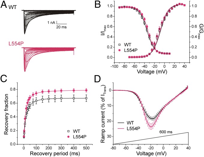
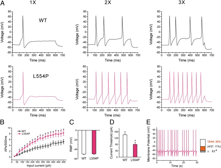
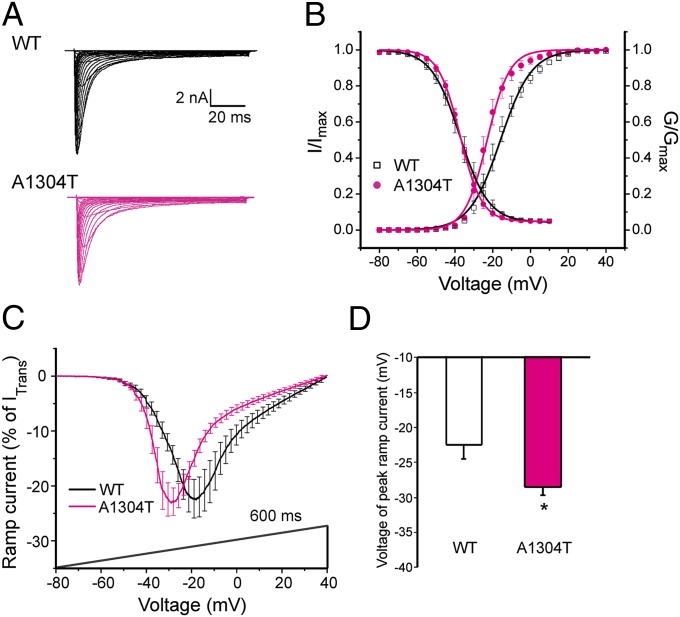
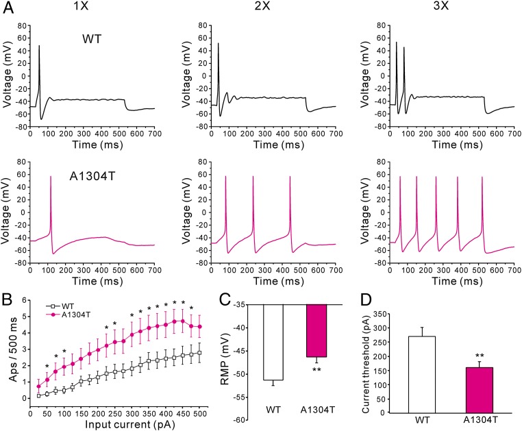
Painful peripheral neuropathy often occurs without apparent underlying cause. Gain-of-function variants of sodium channel Na(v)1.7 have recently been found in ∼30% of cases of idiopathic painful small-fiber neuropathy. Here, we describe mutations in Na(v)1.8, another sodium channel that is specifically expressed in dorsal root ganglion (DRG) neurons and peripheral nerve axons, in patients with painful neuropathy. Seven Na(v)1.8 mutations were identified in 9 subjects within a series of 104 patients with painful predominantly small-fiber neuropathy. Three mutations met criteria for potential pathogenicity based on predictive algorithms and were assessed by voltage and current clamp. Functional profiling showed that two of these three Na(v)1.8 mutations enhance the channel's response to depolarization and produce hyperexcitability in DRG neurons. These observations suggest that mutations of Na(v)1.8 contribute to painful peripheral neuropathy.


Conflict of interest statement

The authors declare no conflict of interest.

Figures

Fig. 1.
Fig. 1.
Loss of IENFs. Immunohistochemical studies (polyclonal anti-protein gene product 9.5 antibody) in sections of skin biopsy at distal leg in a father–son pair (A and B), both harboring L554P mutation in Nav1.8, and in healthy subject (C). Arrows indicate IENF; arrowheads indicate dermal nerve bundles. Patient 2 (A) showed complete depletion of IENFs and severe reduction in density of dermal nerve bundles, which appeared fragmented due to axonal degeneration. Patient 1 (B) showed almost complete loss of IENFs. Inset shows IENF (white arrow) branching after penetrating the epidermis, with large swelling (bent arrow) representing typical axonal predegeneration state. A and B are typical of SFN. For comparison, C shows diffuse innervation of epidermis and dermis in a healthy subject. (Scale bar, 50 μm.)
Fig. 2.
Fig. 2.
L554P mutation alters hNav1.8 channel properties. (A) Representative hNav1.8 WT (black) and L554P (red) currents recorded from DRG neurons. (B) Activation and steady-state fast inactivation were not significantly different. (C) L554P displays enhanced recovery from fast inactivation at −50 mV, close to resting membrane potential of DRG neurons, compared with WT channel (P < 0.05). Data are presented as means ± SEM. (D) Averaged ramp currents (normalized to maximal transient peak current) produced by WT Nav1.8 (black; n = 25) and L554P (red; n = 23) channels. SEM is indicated by error bars every 2 mV. Mean normalized amplitude of peak ramp currents are significantly larger for L554P (WT: 22.3 ± 1.6%, n = 25; L554P: 28.9 ± 2.2%, n = 23; P < 0.05).
Fig. 3.
Fig. 3.
L554P mutation increases the excitability of small DRG neurons. (A) Response of cells expressing WT (Upper) and L554P (Lower) channels, respectively, to 500-ms depolarizing current steps at 1×, 2×, and 3× (left, center, and right traces, respectively) current threshold for action potential generation. (B) Comparison of responses (number of impulses evoked by 500-ms stimulus) in DRG neurons expressing WT (n = 33) and L554P channels (n = 29) across a range of step current injections from 25 to 500 pA (*P < 0.05). (C) Resting membrane potential (RMP) of neurons expressing WT (−50.1 ± 1.3 mV; n = 33) and L554P (−49.6 ± 1.4 mV; n = 29) was not significantly different. (D) Current threshold was significantly reduced after expression of L554P (80 ± 14 pA; n = 29), compared with WT (141 ± 20 pA; n = 33; P < 0.05). (E) Representative recording showing spontaneous firing (30 s) of DRG neuron expressing L554P mutant channels. Trace was recorded for 30 s without current injection. (Inset) Comparison of portions of WT and L554P neurons that produce spontaneous firing.
Fig. 4.
Fig. 4.
A1304T mutation alters hNav1.8 channel properties. (A) Representative hNav1.8 WT (black) and A1304T (red) currents recorded from DRG neurons. (B) Activation for A1304T channels was hyperpolarized by ∼6 mV compared with WT, whereas steady-state fast inactivation of WT and A1304T were not significantly different. (C) Averaged ramp currents (normalized to maximal transient peak current) produced by WT Nav1.8 (black; n = 11) and A1304T (red; n = 12) channels. SEM is indicated by error bars. Voltage dependence of ramp currents for A1304T was hyperpolarized by 6 mV compared with WT. (D) Mean normalized voltage of peak ramp current for A1304T channels (−28.5 ± 1.1 mV; n = 12; *P < 0.05) was significantly different from that for WT channels (−22.5 ± 1.9 mV; n = 11). Data are presented as means ± SEM. *P < 0.05.
Fig. 5.
Fig. 5.
A1304T increases the excitability of small DRG neurons. (A) Response of cells expressing WT (Upper) and A1304T (Lower) channels, respectively, to 500-ms depolarizing current steps that are 1×, 2×, and 3× (left, center, and right traces, respectively) of current threshold for action potential generation. (B) Comparison of responses (number of impulses evoked by a 500-ms stimulus) in DRG neurons expressing WT (n = 34) and A1304T channels (n = 35) across a range of step current injections from 25 to 500 pA; *P < 0.05. (C) Resting membrane potential for A1304T channels was depolarized by 5 mV compared with WT (WT: −51.3 ± 1.2 mV, n = 34; A1304T: −46.3 ± 1.3 mV, n = 35; **P < 0.01). (D) Current threshold to 200-ms stimuli was reduced in DRG neurons expressing A1304T (161 ± 21 pA; n = 35) compared with WT channels (270 ± 32 pA; n = 34; **P < 0.01).

References

    1. Hoeijmakers JG, Faber CG, Lauria G, Merkies IS, Waxman SG. Small-fibre neuropathies—advances in diagnosis, pathophysiology and management. Nat Rev Neurol. 2012;8(7):369–379. - PubMed
    1. Faber CG, et al. Gain of function Naν1.7 mutations in idiopathic small fiber neuropathy. Ann Neurol. 2012;71(1):26–39. - PubMed
    1. Persson AK, et al. Sodium-calcium exchanger and multiple sodium channel isoforms in intra-epidermal nerve terminals. Mol Pain. 2010;6:84. - PMC - PubMed
    1. Lauria G, et al. Intraepidermal nerve fiber density at the distal leg: A worldwide normative reference study. J Peripher Nerv Syst. 2010;15(3):202–207. - PubMed
    1. Reulen JP, Lansbergen MD, Verstraete E, Spaans F. Comparison of thermal threshold tests to assess small nerve fiber function: Limits vs. levels. Clin Neurophysiol. 2003;114(3):556–563. - PubMed

Publication types

Substances