. 2012 Nov 20;109(47):E3268-77.
doi: 10.1073/pnas.1217207109. Epub 2012 Oct 30.

Complex-type N-glycan recognition by potent broadly neutralizing HIV antibodies

Affiliations

Complex-type N-glycan recognition by potent broadly neutralizing HIV antibodies

Hugo Mouquet et al. Proc Natl Acad Sci U S A. .

Abstract

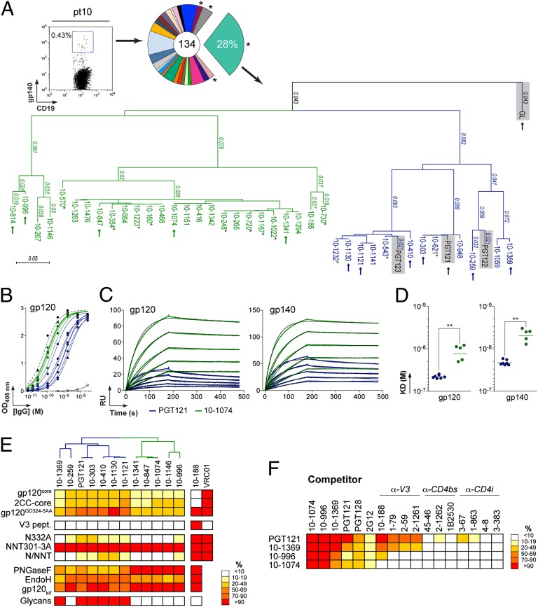
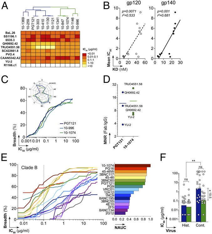
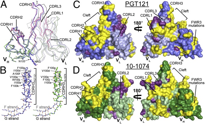
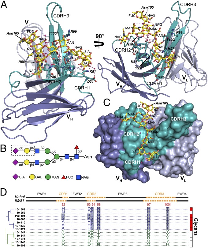
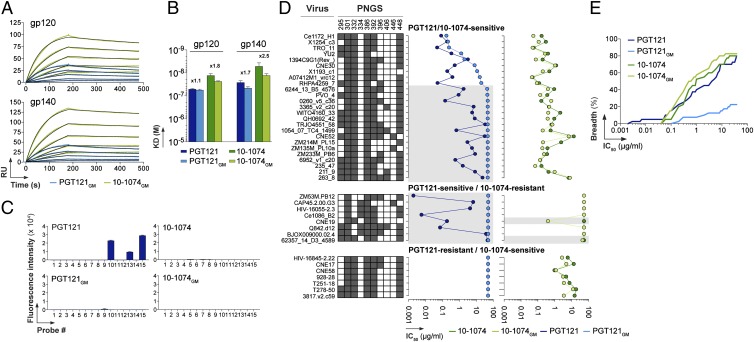
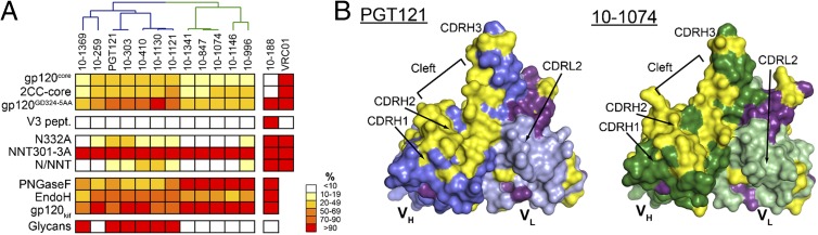
Broadly neutralizing HIV antibodies (bNAbs) can recognize carbohydrate-dependent epitopes on gp120. In contrast to previously characterized glycan-dependent bNAbs that recognize high-mannose N-glycans, PGT121 binds complex-type N-glycans in glycan microarrays. We isolated the B-cell clone encoding PGT121, which segregates into PGT121-like and 10-1074-like groups distinguished by sequence, binding affinity, carbohydrate recognition, and neutralizing activity. Group 10-1074 exhibits remarkable potency and breadth but no detectable binding to protein-free glycans. Crystal structures of unliganded PGT121, 10-1074, and their likely germ-line precursor reveal that differential carbohydrate recognition maps to a cleft between complementarity determining region (CDR)H2 and CDRH3. This cleft was occupied by a complex-type N-glycan in a "liganded" PGT121 structure. Swapping glycan contact residues between PGT121 and 10-1074 confirmed their importance for neutralization. Although PGT121 binds complex-type N-glycans, PGT121 recognized high-mannose-only HIV envelopes in isolation and on virions. As HIV envelopes exhibit varying proportions of high-mannose- and complex-type N-glycans, these results suggest promiscuous carbohydrate interactions, an advantageous adaptation ensuring neutralization of all viruses within a given strain.


Conflict of interest statement

Conflict of interest statement: M.C.N., H.M., P.J.B. and L.S. have a pending patent application for the new PGT121 antibody variants described in the present study with the United States Patent and Trademark Office.

Figures

Fig. 1.
Fig. 1.
PGT121 and 10-1074 clonal variants. (A) (Left) Cytogram showing staining of pt10 PBMCs. (Center) Pie chart showing the expansion of gp140-specific IgG memory B cells. *, no matching IgL was initially found. (Right) Dendrogram showing the relationship between PGT121-like (blue) and 10-1074–like (green) IgH protein sequences with PGT121, PGT122, PGT123, and GL sequences (gray shading) included for comparison. Arrows indicate clones that were produced as IgGs. Underlined clones were not identical at the nucleotide level. *, no matching IgL could be amplified due to limited cDNA material. (B) ELISA comparisons of binding. Curves show the binding of 10-1074–like (green) and PGT121-like (blue) antibodies to YU-2 gp120. Black dashed and solid lines show positive (10-188) and negative (mG053) controls, respectively. (C) SPR sensorgrams showing the binding of 10-1074 (green) and PGT121 (blue) to YU-2 gp120 or gp140. Fits to a 1:1 binding model are shown in black. RU, response units. (D) Apparent KD values for the binding of 10-1074–like (green dots) and PGT121-like (blue dots) antibodies to gp120 and gp140. **P < 0.005. (E) Heat map (expressed as percentage of binding to unmodified gp120) summarizing the binding of PGT121-like and 10-1074–like antibodies [listed with a dendrogram showing their relationships (A)] to diverse antigens (SI Appendix, Figs. S3–S8). Darker colors, stronger binding; white, no observed binding. Anti-gp120V3 (10-188) and anti-CD4bs (VRC01) antibodies are controls. For V3 loop peptide-binding assays (V3 pept.) and glycan arrays, red indicates binding and white indicates no binding. (F) Competition ELISAs. Heat map showing the relative binding to gp120 of selected PGT121-like and 10-1074–like antibodies in the presence of potential competitor antibodies (SI Appendix, Fig. S8). Results are expressed as percentage of binding in presence of 100 µg/mL of competitor compared with binding in the absence of competitor. Darker colors indicate stronger inhibition; white indicates no competition.
Fig. 2.
Fig. 2.
Neutralization activity of PGT121-like and 10-1074–like variants. (A) Heat map comparing the neutralization potencies of PGT121-like and 10-1074–like antibodies (listed at the top with a dendrogram showing relationships; Fig. 1A) in the TZM-bl assay. Darker colors, more potent neutralization; white, no neutralization. (B) Correlation between the mean IC80 against nine viruses (y axis) and apparent KD values for binding to gp120 and gp140 (x axis). (C) Graph comparing the neutralization breadth and potencies of PGT121, 10-996, and 10-1074 antibodies in the TZM-bl assay against an extended panel of 119 viruses. The y axis shows the cumulative frequency of IC50 values up to the concentration shown on the x axis. The spider graph (upper left) shows the frequency distribution of neutralized viruses according to HIV-1 clades. (D) Dot plot showing molar neutralization ratios (MNRs) (ratio of the Fab and IgG IC50 concentrations). Horizontal bars represent the mean IC50s for all viruses. (E) Coverage graph (as in C) comparing the neutralization breadth and potencies of selected bNAbs against a panel of 95 clade B viruses as evaluated by the PBMC-based neutralization assay. (Right) Bar graph showing values for the normalized area under the curve (NAUC) for the IgGs shown in the coverage graph. (F) Bar graph comparing the neutralization potencies of PGT121 (blue) and 10-1074 (green) against viruses isolated from historical (Hist.) and contemporary (Cont.) seroconverters. ns, nonsignificant; **P < 0.005. Fold difference between median IC50s for the neutralization of contemporary viruses by PGT121 and 10-1074 is indicated.
Fig. 3.
Fig. 3.
Comparison of PGT121, 10-1074, and GL Fab structures. (A) Cα alignment of variable domains of PGT121 (VHPGT121, blue; VLPGT121, light blue), 10-1074 (VH10-1074, green; VL10-1074, light green) and GL (VHGL, dark gray; VLGL, light gray) (CDRs for 10-1074 are magenta). An N-acetylglucosamine attached to PGT121 Asn105HC is depicted as sticks. (B) CDRH3 loops of PGT121 and 10-1074. Extensions of the VH domain F (white) and G (grey) strands (only main chain atoms shown) form CDRH3, a two-stranded β-sheet in PGT121 (blue), 10-1074 (green), and GL (main and side chain atoms shown; main chain hydrogen bonds are yellow dashes). (C and D) Surface representation of PGT121 (C) and 10-1074 (D) variable domains showing differences (yellow) and somatic mutations in common (purple). PGT121 HC, blue; PGT121 LC, light blue; 10-1074 HC, green; 10-1074 LC, light green.
Fig. 4.
Fig. 4.
Crystal structure of a liganded PGT121. (A) Ribbon diagram of the PGT121 variable domains bound to a complex-type N-glycan (yellow sticks) attached to Asn105HC of a neighboring Fab in the crystal. VH is blue with CDRs highlighted in teal, VL is light blue, side-chain and backbone atoms of contact residues are depicted as sticks, hydrogen bonds are magenta dashed lines, and water molecules are red spheres. (B) Schematic of the bound complex-type glycan. Glycan residues in the dashed box were disordered. Fucose (FUC), red triangles; galactose (GAL), yellow circles; mannose (MAN), green circles; N-acetyl glucosamine (NAG), blue squares; sialic acid (SIA), magenta diamonds. (C) Surface representation of PGT121 variable domains with bound glycan (sticks). Coloring is as in A. (D) Amino acid alignment comparing residues at positions 32, 53, 54, 58, 97, and 100l (residues of PGT121 making direct or likely contacts with N-glycan; amino acid numbering based on crystal structures) between PGT121-like (blue) and 10-1074–like (green) antibodies. Framework (FWR) and complementarity determining regions (CDR) (Upper), dendrogram showing relationships (Left), and binding to protein-free glycans as detected in glycan arrays (Right; red indicates binding, white indicates no binding) are indicated. Gray shading indicates amino acid identity.
Fig. 5.
Fig. 5.
Binding and neutralization activities of PGT121GM and 10-1074GM mutant antibodies. (A) SPR sensorgrams showing the binding of PGT121GM (light green) and 10-1074GM (light blue) to YU-2 gp120 or gp140. (B) Bar graphs comparing apparent KD values for the binding of 10-1074, PGT121, PGT121GM, and 10-1074GM antibodies to gp120 and gp140. Error bars indicate the SEM of KD values obtained from three independent experiments. Fold differences between KD values of wild-type vs. glycomutant antibodies are indicated. (C) Bar graphs comparing binding of glycans (SI Appendix, Fig. S6A) by PGT121 and 10-1074 with mutant antibodies (PGT121GM and 10-1074GM). Numerical scores of binding are measured as fluorescence intensity (means at duplicate spots) for probes arrayed at 5 fmol/spot. (D) Graphs comparing IC50 values for neutralization of selected viruses by PGT121, 10-1074, PGT121GM, and 10-1074GM in the TZM-bl assay. Viral strains are listed with PNGSs at the indicated gp120 positions (those near Asn332gp120) color coded: dark gray, a PNGS; white, not a PNGS. From the 2012 Los Alamos filtered web alignment of ∼2,865 gp120 sequences, the frequencies of PNGSs at these positions are 295 (58.3%), 301 (91.7%), 332 (73.0%), 334 (20.4%), 386 (86.7%), 392 (79.4%), 396 (25.7%), 406 (5.5%), 446 (7.6%), and 448 (85.6%). Gray shading indicates PGT121-sensitive viruses that were not neutralized by PGT121GM and a 10-1074–resistant strain that was neutralized by 10-1074GM. (E) Coverage graph comparing the neutralization breadth and potencies of PGT121, PGT121GM, 10-1074, and 10–1074GM antibodies in the TZM-bl assay against a panel of 40 viruses.
Fig. P1.
Fig. P1.
(A) Heat map (expressed as percentage of binding to unmodified gp120) summarizing the binding of PGT121-like and 10-1074–like antibodies to antigens. Darker colors represent stronger binding; white represents no detectable binding. A dendrogram above the antibody names divides them into PGT121-like (blue lines) and 10-1074–like (green lines) antibodies. Antibodies against the gp120 V3 loop (10-188) and CD4-binding site (VRC01) are controls. For V3 loop peptide-binding assays (V3 pept.) and glycan microarrays (Glycans), red indicates binding and white indicates no binding. PNGase F and Endo H are glycosidase enzymes that can cleave complex-type and high-mannose glycans (PNGase F) or high-mannose glycans (Endo H). N332A indicates the substitution of asparagine at gp120 position 332 to alanine. (B) Surface representation of PGT121 (blue) and 10-1074 (green) variable domain structures showing the location of differences (yellow) and somatic mutations in common (purple).

References

    1. Plotkin SA. Vaccines: Correlates of vaccine-induced immunity. Clin Infect Dis. 2008;47(3):401–409. - PubMed
    1. Haynes BF, et al. Immune-correlates analysis of an HIV-1 vaccine efficacy trial. N Engl J Med. 2012;366(14):1275–1286. - PMC - PubMed
    1. Simek MD, et al. Human immunodeficiency virus type 1 elite neutralizers: Individuals with broad and potent neutralizing activity identified by using a high-throughput neutralization assay together with an analytical selection algorithm. J Virol. 2009;83(14):7337–7348. - PMC - PubMed
    1. Sather DN, et al. Factors associated with the development of cross-reactive neutralizing antibodies during human immunodeficiency virus type 1 infection. J Virol. 2009;83(2):757–769. - PMC - PubMed
    1. Gray ES, et al. Antibody specificities associated with neutralization breadth in plasma from human immunodeficiency virus type 1 subtype C-infected blood donors. J Virol. 2009;83(17):8925–8937. - PMC - PubMed

Publication types

MeSH terms