

Knowledge building insights on biomarkers of arsenic toxicity to keratinocytes and melanocytes
- PMID: 23115478
- PMCID: PMC3480875
- DOI: 10.4137/BMI.S7799
Knowledge building insights on biomarkers of arsenic toxicity to keratinocytes and melanocytes
Abstract
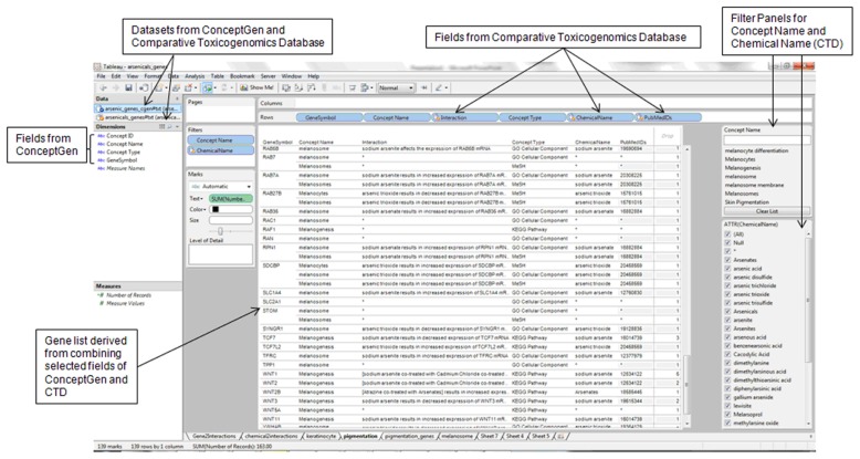
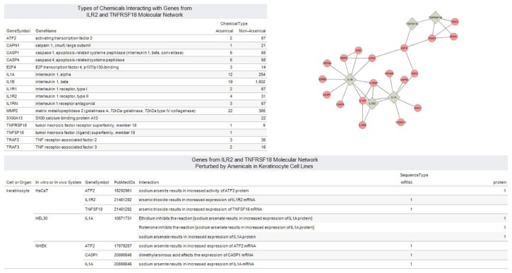
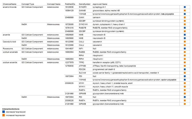
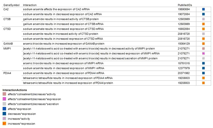
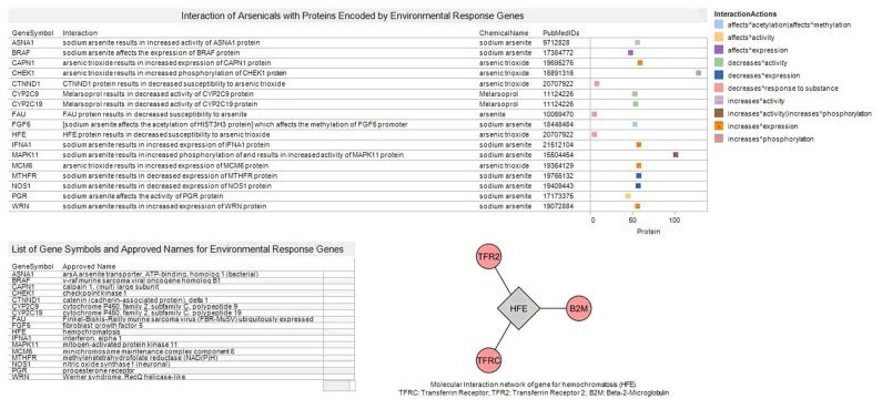
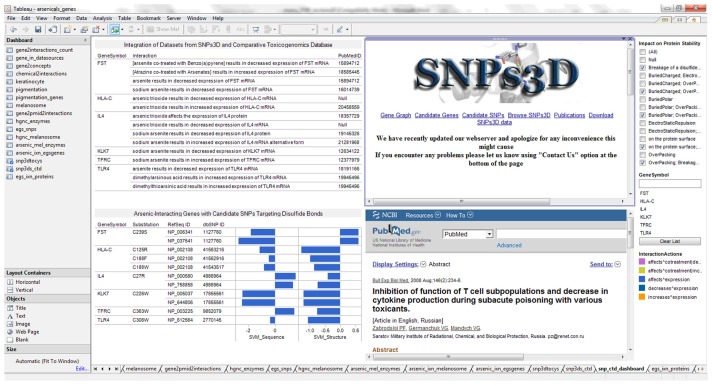
Exposure to inorganic arsenic induces skin cancer and abnormal pigmentation in susceptible humans. High-throughput gene transcription assays such as DNA microarrays allow for the identification of biological pathways affected by arsenic that lead to initiation and progression of skin cancer and abnormal pigmentation. The overall purpose of the reported research was to determine knowledge building insights on biomarker genes for arsenic toxicity to human epidermal cells by integrating a collection of gene lists annotated with biological information. The information sets included toxicogenomics gene-chemical interaction; enzymes encoded in the human genome; enriched biological information associated with genes; environmentally relevant gene sequence variation; and effects of non-synonymous single nucleotide polymorphisms (SNPs) on protein function. Molecular network construction for arsenic upregulated genes TNFSF18 (tumor necrosis factor [ligand] superfamily member 18) and IL1R2 (interleukin 1 Receptor, type 2) revealed subnetwork interconnections to E2F4, an oncogenic transcription factor, predominantly expressed at the onset of keratinocyte differentiation. Visual analytics integration of gene information sources helped identify RAC1, a GTP binding protein, and TFRC, an iron uptake protein as prioritized arsenic-perturbed protein targets for biological processes leading to skin hyperpigmentation. RAC1 regulates the formation of dendrites that transfer melanin from melanocytes to neighboring keratinocytes. Increased melanocyte dendricity is correlated with hyperpigmentation. TFRC is a key determinant of the amount and location of iron in the epidermis. Aberrant TFRC expression could impair cutaneous iron metabolism leading to abnormal pigmentation seen in some humans exposed to arsenicals. The reported findings contribute to insights on how arsenic could impair the function of genes and biological pathways in epidermal cells. Finally, we developed visual analytics resources to facilitate further exploration of the information and knowledge building insights on arsenic toxicity to human epidermal keratinocytes and melanocytes.
Keywords: RAC1; SNPs; TFRC; arsenic; disulfide bond; environmental response genes; functional annotation; hyperpigmentation; iron uptake; keratinocyte; melanocyte; skin cancer; toxicogenomics; vicinal cysteines Isokpehi et al.
Figures
Similar articles
-
Proteomics-Based Identification of Differentially Abundant Proteins from Human Keratinocytes Exposed to Arsenic Trioxide.J Proteomics Bioinform. 2014 Jul;7(7):166-178. doi: 10.4172/jpb.1000317. J Proteomics Bioinform. 2014. PMID: 25419056 Free PMC article.
-
Aberrantly Expressed Genes in HaCaT Keratinocytes Chronically Exposed to Arsenic Trioxide.Biomark Insights. 2011 Feb 8;6:7-16. doi: 10.4137/BMI.S6383. Biomark Insights. 2011. PMID: 21461292 Free PMC article.
-
Candidate single nucleotide polymorphism markers for arsenic responsiveness of protein targets.Bioinform Biol Insights. 2010 Oct 11;4:99-111. doi: 10.4137/BBI.S5498. Bioinform Biol Insights. 2010. PMID: 20981267 Free PMC article.
-
[Culture of human melanocytes. Its contribution to the knowledge of melanocyte physiology].Pathol Biol (Paris). 1992 Feb;40(2):114-20. Pathol Biol (Paris). 1992. PMID: 1608652 Review. French.
-
Precise role of dermal fibroblasts on melanocyte pigmentation.J Dermatol Sci. 2017 Nov;88(2):159-166. doi: 10.1016/j.jdermsci.2017.06.018. Epub 2017 Jul 1. J Dermatol Sci. 2017. PMID: 28711237 Review.
Cited by
-
MiR-96-5p Suppresses Progression of Arsenite-Induced Human Keratinocyte Proliferation and Malignant Transformation by Targeting Denticleless E3 Ubiquitin Protein Ligase Homolog.Toxics. 2023 Dec 1;11(12):978. doi: 10.3390/toxics11120978. Toxics. 2023. PMID: 38133379 Free PMC article.
-
State of the science review of the health effects of inorganic arsenic: Perspectives for future research.Environ Toxicol. 2019 Feb;34(2):188-202. doi: 10.1002/tox.22673. Epub 2018 Dec 4. Environ Toxicol. 2019. PMID: 30511785 Free PMC article. Review.
-
Melanocytotoxic chemicals and their toxic mechanisms.Toxicol Res. 2022 Aug 22;38(4):417-435. doi: 10.1007/s43188-022-00144-2. eCollection 2022 Oct. Toxicol Res. 2022. PMID: 36277364 Free PMC article. Review.
-
Proteomics-Based Identification of Differentially Abundant Proteins from Human Keratinocytes Exposed to Arsenic Trioxide.J Proteomics Bioinform. 2014 Jul;7(7):166-178. doi: 10.4172/jpb.1000317. J Proteomics Bioinform. 2014. PMID: 25419056 Free PMC article.
-
Secondary Data Analytics of Aquaporin Expression Levels in Glioblastoma Stem-Like Cells.Cancer Inform. 2015 Jul 30;14:95-103. doi: 10.4137/CIN.S22058. eCollection 2015. Cancer Inform. 2015. PMID: 26279619 Free PMC article.
References
-
- Hall M, Chen Y, Ahsan H, et al. Blood arsenic as a biomarker of arsenic exposure: results from a prospective study. Toxicology. 2006;225:225–33. - PubMed
-
- Tchounwou PB, Patlolla AK, Centeno JA. Carcinogenic and systemic health effects associated with arsenic exposure—a critical review. Toxicol Pathol. 2003;31:575–88. - PubMed
-
- Garelick H, Jones H, Dybowska A, Valsami-Jones E. Arsenic pollution sources. Rev Environ Contam Toxicol. 2008;197:17–60. - PubMed
Grants and funding
LinkOut - more resources
Full Text Sources
Research Materials
Miscellaneous
