. 2011 Oct 13;4(10):1328-1354.
doi: 10.3390/ph4101328.

Targeting the Large Subunit of Human Ribonucleotide Reductase for Cancer Chemotherapy

Affiliations

Targeting the Large Subunit of Human Ribonucleotide Reductase for Cancer Chemotherapy

Sanath R Wijerathna et al. Pharmaceuticals (Basel). .

Abstract

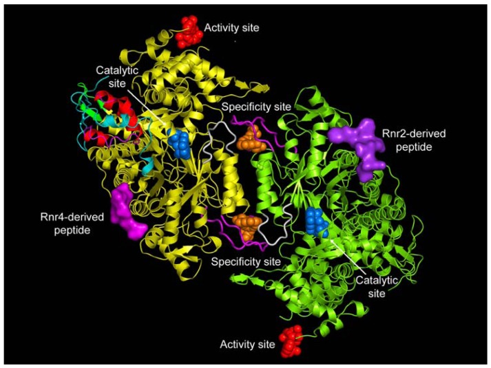
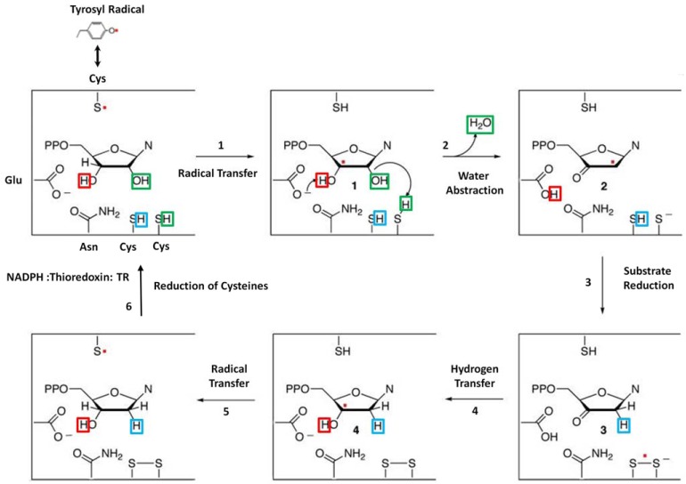
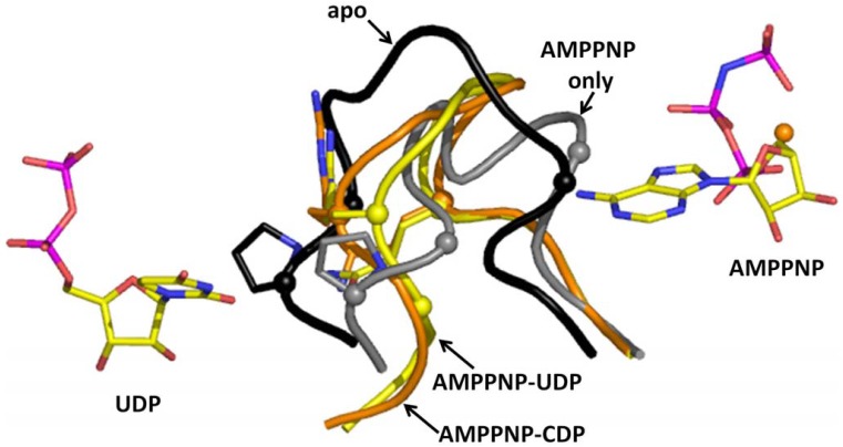
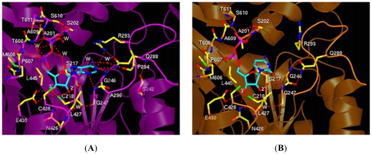
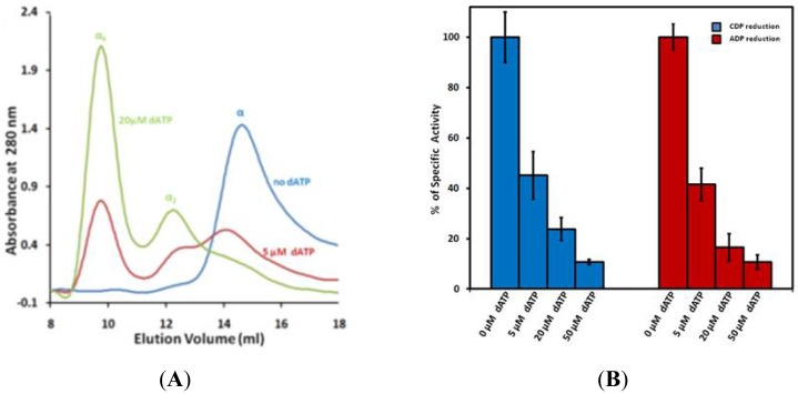
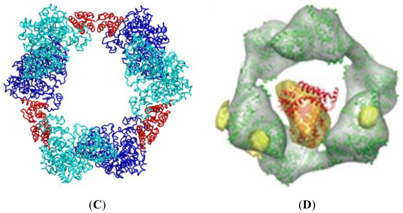
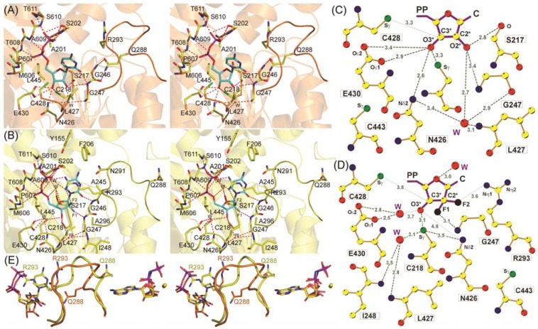
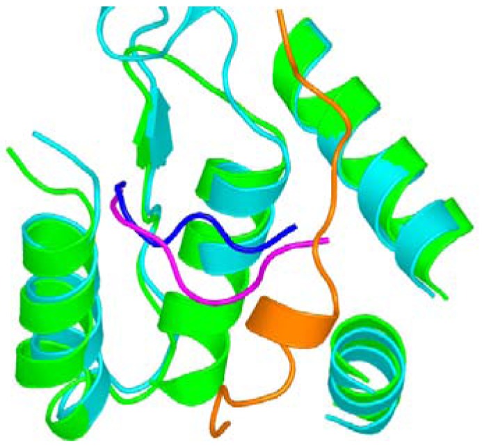
Ribonucleotide reductase (RR) is a crucial enzyme in de novo DNA synthesis, where it catalyses the rate determining step of dNTP synthesis. RRs consist of a large subunit called RR1 (α), that contains two allosteric sites and one catalytic site, and a small subunit called RR2 (β), which houses a tyrosyl free radical essential for initiating catalysis. The active form of mammalian RR is an α(n)β(m) hetero oligomer. RR inhibitors are cytotoxic to proliferating cancer cells. In this brief review we will discuss the three classes of RR, the catalytic mechanism of RR, the regulation of the dNTP pool, the substrate selection, the allosteric activation, inactivation by ATP and dATP, and the nucleoside drugs that target RR. We will also discuss possible strategies for developing a new class of drugs that disrupts the RR assembly.


Figures

Figure 1
Figure 1
Class I Ribonucleotide reductase I. The specificity (S-site), catalytic (C-site), and activity (A-site) are shown as solid objects. Rnr2/Rnr4 peptides define the P-site. Loop 2 (white) and loop 1 (magenta) are close to the dimer interface. Reproduced with permission from PNAS [12]. Copyright (2006) National Academy of Sciences, USA.
Figure 2
Figure 2
Catalytic Mechanism of Class Ia RR. See the text for a detailed description of the mechanism. Adopted from Zipse et al. [51] and modified. Copyright (2009) American Chemical Society.
Figure 3
Figure 3
Loop 2 movements upon binding of effectors: Substrate (CDP) and loop 2 and effector (AMPPNP) are shown for AMPPNP-UDP. Loop 2 is shown for apo (black), AMPPNP only (gray), and AMPPNP-CDP(orange). Reproduced with permission from PNAS [13]. Copyright (2006) National Academy of Sciences, USA.
Figure 4
Figure 4
Substrate selection. (A) ADP binding at the C-site and (B) CDP binding at the C-site. The key residues on loop 2 required for substrate selection are Q288 and R293 are to the right. The catalytic residues C218, C428, N426 and E428 are also shown binding to the ribose moiety. Reproduced with permission from PNAS [13]. Copyright (2006) National Academy of Sciences, USA.
Figure 5
Figure 5
Characterization of dATP hexamer: (A) human RR1(hRR1) was tested for its ability to form hexamers in the presence of varying concentrations of dATP. No oligomers were observed in the absence of dATP (blue trace) and a mixed population of monomers, dimers, and hexamers at a dATP concentration of 5 μM (red trace). At 20 μM dATP, the hexamers are the dominant species, with a small amount of dimer (green trace). (B) The specific activity of the wild type enzyme decreased with increasing concentration of dATP. Activities for [1H] CDP reduction (blue) and [14C] ADP (red) reduction is shown. (C) Hexameric packing of RR1 based on the low-resolution X-ray crystal structure of the ScRR1 hexamer. ScRR1 monomers are colored in forest green and limon or blue and cyan. All the four-helix ATP-binding cones are colored in red. (D) Model of the α6●ββ′●dATP holo complex Reproduced with the permission from NSMB 2011 [26].
Figure 5
Figure 5
Characterization of dATP hexamer: (A) human RR1(hRR1) was tested for its ability to form hexamers in the presence of varying concentrations of dATP. No oligomers were observed in the absence of dATP (blue trace) and a mixed population of monomers, dimers, and hexamers at a dATP concentration of 5 μM (red trace). At 20 μM dATP, the hexamers are the dominant species, with a small amount of dimer (green trace). (B) The specific activity of the wild type enzyme decreased with increasing concentration of dATP. Activities for [1H] CDP reduction (blue) and [14C] ADP (red) reduction is shown. (C) Hexameric packing of RR1 based on the low-resolution X-ray crystal structure of the ScRR1 hexamer. ScRR1 monomers are colored in forest green and limon or blue and cyan. All the four-helix ATP-binding cones are colored in red. (D) Model of the α6●ββ′●dATP holo complex Reproduced with the permission from NSMB 2011 [26].
Figure 6
Figure 6
Catalytic site interactions of CDP and F2dCDP taken from Xu et al., PNAS 2006. (A) Stereo view of CDP (orange). Interacting atoms: oxygen, red; nitrogen, blue; phosphate, magenta; sulfur green; substrate carbons, cyan; protein non-Cα, yellow; Cα, as secondary structure, orange. (B) Stereo view of F2dCDP. Interacting atoms are colored as in (A) above except sulfur orange; Cα carbons, as secondary structure, yellow; fluorines, grey. (C) Ligand plot of CDP ribose interactions. Colors are as in Figure 6(A), except that carbons are yellow and fluorine, black. (D) Ligand plot of F2dCDP interactions. The van der Waals contact to L427 is omitted for clarity. (E) Stereo view of loop 2 superposition of AMPPNP-CDP (orange) and AMPPNP-F2dCDP (yellow). Substrate/inhibitor is seen on the left and the effector is on the right. Reproduced with permission from PNAS. Copyright (2006) National Academy of Sciences, USA [84].
Figure 7
Figure 7
View of the structural comparison of ScRR2, ScRR4 and EcRR2 peptide binding. Carbon atoms for ScRR2 peptide (magenta), ScRR4 peptide (blue) and EcRR2 peptide (orange). Nearby helices are drawn from the ScRR1-ScRR2 complex (green) and EcRR1-EcRR2 complex (cyan). The figure was reproduced with permission from PNAS [12]. Copyright (2006) National Academy of Sciences, USA.
Figure 8
Figure 8
Binding of P7 to ScRR1. (A) P7 binds at subsites 1 and 2 connected by a reverse turn (B) P7-yellow where subsite 1 is to the right involving F1 and subsite 2 is to the left involving D6F7. ScRR1 binding site is depicted as a surface. The figure was reproduced with permission from Xu et al. [97]. Copyright (2008) American Chemical Society.
Figure 9
Figure 9
P6 binding to ScRR1. P6-orange, The figure was reproduced with permission from Xu et al., [97]. Copyright (2008) American Chemical Society.
Figure 10
Figure 10
Cyc 10 (A) Cyc 10 structure (B) Mode of binding of Cyc 10 to ScRR1.

References

    1. Nordlund P., Reichard P. Ribonucleotide reductases. Annu. Rev. Biochem. 2006;75:681–706. - PubMed
    1. Kolberg M., Strand K.R., Graff P., Andersson K.K. Structure, function, and mechanism of ribonucleotide reductases. Biochim. Biophys. Acta. 2004;1699:1–34. - PubMed
    1. Bertani L.E., Haggmark A., Reichard P. Synthesis of pyrimidine deoxyribonucleoside diphosphates with enzymes from Escherichia coli. J. Biol. Chem. 1961;236:PC67–PC68. - PubMed
    1. Jordan A., Reichard P. Ribonucleotide reductases. Annu. Rev. Biochem. 1998;67:71–98. - PubMed
    1. Reichard P. From RNA to DNA, why so many ribonucleotide reductases? Science. 1993;260:1773–1777. - PubMed

LinkOut - more resources