. 2012 Nov 1:11:82.
doi: 10.1186/1476-4598-11-82.

The DEAD/DEAH box helicase, DDX11, is essential for the survival of advanced melanomas

Affiliations

The DEAD/DEAH box helicase, DDX11, is essential for the survival of advanced melanomas

Chitralekha Bhattacharya et al. Mol Cancer. .

Abstract

Background: Despite continuous efforts to identify genes that are pivotal regulators of advanced melanoma and closely related to it, to determine which of these genes have to be blocked in their function to keep this highly aggressive disease in check, it is far from clear which molecular pathway(s) and specific genes therein, is the Achilles' heel of primary and metastatic melanoma. In this report, we present data, which document that the DEAD-box helicase DDX11, which is required for sister chromatid cohesion, is a crucial gatekeeper for melanoma cell survival.

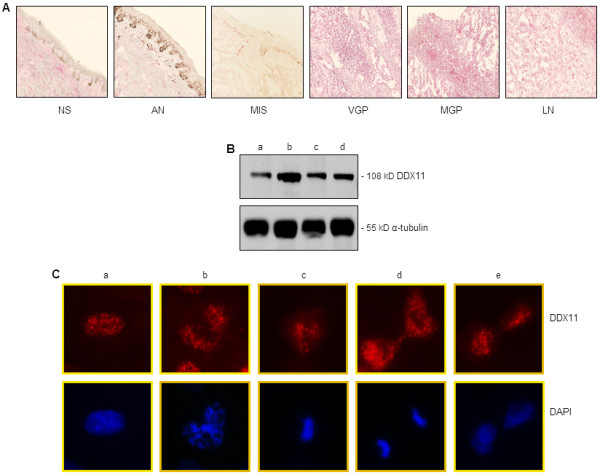
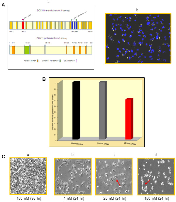
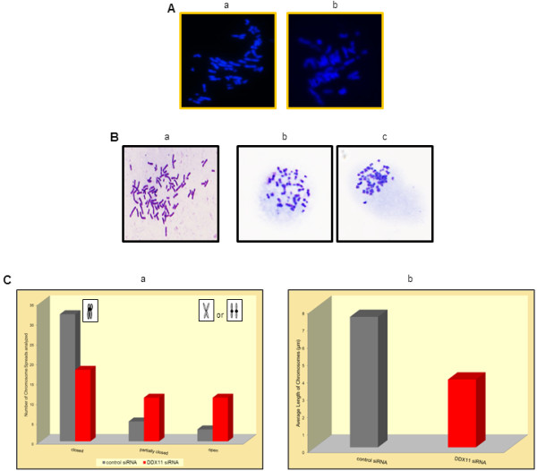
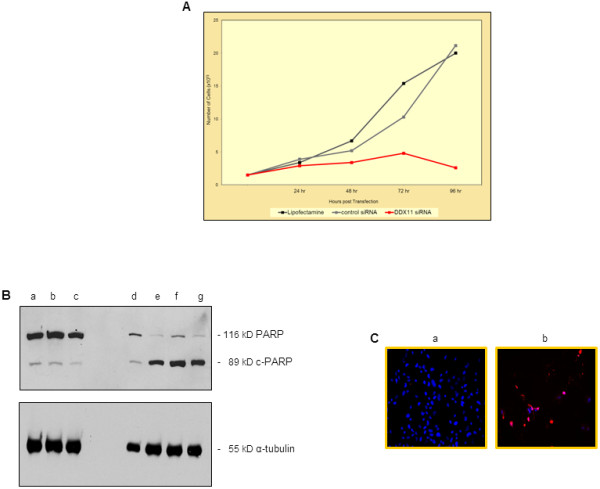
Methods: Performing immunohistochemistry and immunoblot analysis, we determined expression of DDX11 in melanoma tissues and cell lines. Following transfection of melanoma cells with a DDX11-specific siRNA, we conducted a qPCR analysis to determine downregulation of DDX11 in the transfected melanoma cells. In subsequent studies, which focused upon an analysis of fluorescently labeled as well as Giesma-stained chromosome spreads, a proliferation analysis and apoptosis assays, we determined the impact of suppressing DDX11 expression on melanoma cells representing advanced melanoma.

Result: The findings of the study presented herein document that DDX11 is upregulated with progression from noninvasive to invasive melanoma, and that it is expressed at high levels in advanced melanoma. Furthermore, and equally important, we demonstrate that blocking the expression of DDX11 leads not only to inhibition of melanoma cell proliferation and severe defects in chromosome segregation, but also drives melanoma cells rapidly into massive apoptosis.

Conclusion: To date, little is known as to whether helicases play a role in melanoma development and specifically, in the progression from early to advanced melanoma. In this report, we show that the helicase DDX11 is expressed at high levels in primary and metastatic melanoma, and that interfering with its expression leads to severe chromosome segregation defects, telomere shortening, and massive melanoma cell apoptosis. These findings suggest that DDX11 could be an important candidate for molecular targeted therapy for advanced melanoma.


Figures

Figure 1
Figure 1
DDX11 expression in normal skin, nevi, and melanoma tissues and cell lines. (A) Cryopreserved tissue sections, representing normal human skin (NS), atypical nevus (AN), melanoma in situ (MIS), VGP melanoma (VGP), MGP melanoma (MGP), and melanoma-infiltrated lymph node (LN), were stained with an antibody to human DDX11 and counterstained with hematoxylin. Images were captured at 10X magnification. (B) Immunoblot analysis of VGP melanoma cells WM983-A (a), and MGP melanoma cells WM983-B (b), WM852 (c), and WM1158 (d) probed with DDX11 antibody. For loading control, the immunoblot was probed with an antibody to α-tubulin. (C) Immunofluorescence analysis of DDX11 expression in MGP melanoma cells, WM1158, during interphase (a), prophase (b), metaphase (c), telophase (d) and late telophase (e). The melanoma cells were probed with an antibody to human DDX11 (pseudocolored red), and their nuclei (pseudocolored blue) counterstained with fluorescent DAPI.
Figure 2
Figure 2
Analysis of DDX11 siRNA-transfected melanoma cells. (A, panel a) Schematic presentation of human DDX11 transcript variant 1, and protein isoform 1. The red-colored symbol above exon 3 in transcript 1 marks the position of the DDX11 siRNA, and the green-colored bar symbol above exons 20–22, indicates the location of the DDX11 qPCR primers used in the qPCR analysis. (A, panel b) Immunofluorescence analysis of WM1158 MGP melanoma cells at 24 hr following transfection with 5 nM of Cy5-conjugated DDX11 siRNA (pseudocolored yellow). Nuclei (pseudocolored blue) were counterstained with fluorescent DAPI. (B) qPCR analysis of WM1158 MGP melanoma cells that had received only Lipofectamine 2000 (black-colored bar), or were transfected with 25 nM of control siRNAs (grey-colored bar) or DDX11 siRNA (red-colored bar) for 48 hr. (C) Phase-contrast images, captured at 10X magnification, depicting the morphology WM1158 MGP melanoma cells following transfection with 150 nM of control siRNAs for 96 hr (a) or with 1 nM (b), 25 nM (c) or 150 nM (d) of DDX11 siRNA for 24 hr. The red-colored arrow in the phase-contrast images, shown in panels c and d, points to the chain-like morphology of melanoma cells that did not separate.
Figure 3
Figure 3
Inhibition of DDX11 expression in melanoma cells leads to chromosome segregation defects. (A) Immunofluorescence analysis of fluorescent DAPI-stained chromosome spreads (40X magnification) of WM1158 MGP melanoma cells transfected for 24 hr with 75 nM of control siRNA (a) or 75 nM of DDX11 siRNA (b). (B) Images of Giemsa-stained chromosome spreads of WM1158 MGP melanoma cells transfected for 24 hr with 75 nM of control siRNAs (a) or 75 nM of DDX11 siRNA (b and c). (C) Giemsa-stained chromosome spreads of WM1158 MGP melanoma cells transfected for 24 hr with 75 nM of control siRNAs (grey-colored bars) or 75 nM of DDX11 siRNA (red-colored bars) were analyzed for chromosomes with closed, partially closed, or open/separated arms (panel a) as well as average length of chromosomes (panel b).
Figure 4
Figure 4
Suppressing DDX11 expression in melanoma cells leads to inhibition of melanoma cell proliferation and massive melanoma cell apoptosis. (A) Proliferation of WM1158 MGP melanoma cells that received only Lipofectamine 2000 (black-colored line) or were transfected with 25 nM of control siRNAs (grey-colored line) or 25 nM of DDX11 siRNA (red-colored line). Depicted at each time point following siRNA transfection is the mean of duplicate samples analyzed. (B) Immunoblot analysis of WM1158 MGP melanoma cells, transfected with 25 nM of DDX11 siRNA for 24 hr (d), 48 hr (e), 72 hr (f) or 96 hr (g), were probed with an antibody to c-PARP. Protein lysates prepared at 96 hr from WM1158 cells that were not transfected (a), had received only Lipofectamine 2000 (b), or were transfected with 25 nM of control siRNAs (c) served as controls. For loading control, the immunoblot was probed with an antibody to α-tubulin. (C) WM1158 MGP melanoma cells, transfected for 48 hr with 75 nM of control siRNAs (a) or 75 nM of DDX11 siRNA (b) were analyzed by immunofluorescence-based TUNEL. Melanoma cells that had undergone apoptosis are pseudocolored red (TUNEL staining) and fluorescent DAPI-counterstained nuclei are pseudocolored blue.

References

    1. Berger MF, Levin JZ, Vijayendran K, Sivachenko A, Adiconis X, Maguire J, Johnson LA, Robinson J, Verhaak RG, Sougnez C. et al.Integrative analysis of the melanoma transcriptome. Genome Res. 2010;20:413–427. doi: 10.1101/gr.103697.109. - DOI - PMC - PubMed
    1. Wei X, Walia V, Lin JC, Teer JK, Prickett TD, Gartner J, Stemke-Hale K, Davies MA, Gershenwald JE. NISC Comparative Sequencing Program et al.Exome sequencing identifies GRIN2A as frequently mutated in melanoma. Nat Genet. 2011;43:442–446. doi: 10.1038/ng.810. - DOI - PMC - PubMed
    1. Berger MF, Hodis E, Heffernan TP, Deribe YL, Lawrence MS, Protopopov A, Ivanova E, Watson IR, Nickerson E, Ghosh P. et al.Melanoma genome sequencing reveals frequent PREX2 mutations. Nature. 2012;485:502–506. - PMC - PubMed
    1. Frank S, Werner S. The human homologue of the yeast CHL1 gene is a novel keratinocyte growth factor-regulated gene. J Biol Chem. 1996;271:24337–24340. doi: 10.1074/jbc.271.40.24337. - DOI - PubMed
    1. Amann J, Kidd VJ, Lahti JM. Characterization of putative human homologues of the yeast chromosome transmission fidelity gene, CHL1. J Biol Chem. 1997;272:3823–3832. doi: 10.1074/jbc.272.6.3823. - DOI - PubMed

Publication types

MeSH terms