

Brain region-specific altered expression and association of mitochondria-related genes in autism
- PMID: 23116158
- PMCID: PMC3528421
- DOI: 10.1186/2040-2392-3-12
Brain region-specific altered expression and association of mitochondria-related genes in autism
Abstract
Background: Mitochondrial dysfunction (MtD) has been observed in approximately five percent of children with autism spectrum disorders (ASD). MtD could impair highly energy-dependent processes such as neurodevelopment, thereby contributing to autism. Most of the previous studies of MtD in autism have been restricted to the biomarkers of energy metabolism, while most of the genetic studies have been based on mutations in the mitochondrial DNA (mtDNA). Despite the mtDNA, most of the proteins essential for mitochondrial replication and function are encoded by the genomic DNA; so far, there have been very few studies of those genes. Therefore, we carried out a detailed study involving gene expression and genetic association studies of genes related to diverse mitochondrial functions.
Methods: For gene expression analysis, postmortem brain tissues (anterior cingulate gyrus (ACG), motor cortex (MC) and thalamus (THL)) from autism patients (n=8) and controls (n=10) were obtained from the Autism Tissue Program (Princeton, NJ, USA). Quantitative real-time PCR arrays were used to quantify the expression of 84 genes related to diverse functions of mitochondria, including biogenesis, transport, translocation and apoptosis. We used the delta delta Ct (∆∆Ct) method for quantification of gene expression. DNA samples from 841 Caucasian and 188 Japanese families were used in the association study of genes selected from the gene expression analysis. FBAT was used to examine genetic association with autism.
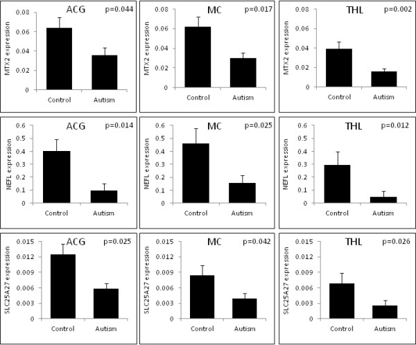
Results: Several genes showed brain region-specific expression alterations in autism patients compared to controls. Metaxin 2 (MTX2), neurofilament, light polypeptide (NEFL) and solute carrier family 25, member 27 (SLC25A27) showed consistently reduced expression in the ACG, MC and THL of autism patients. NEFL (P = 0.038; Z-score 2.066) and SLC25A27 (P = 0.046; Z-score 1.990) showed genetic association with autism in Caucasian and Japanese samples, respectively. The expression of DNAJC19, DNM1L, LRPPRC, SLC25A12, SLC25A14, SLC25A24 and TOMM20 were reduced in at least two of the brain regions of autism patients.
Conclusions: Our study, though preliminary, brings to light some new genes associated with MtD in autism. If MtD is detected in early stages, treatment strategies aimed at reducing its impact may be adopted.
Figures
References
-
- Rice C. Prevalence of autism spectrum disorders-Autism and Developmental Disabilities Monitoring Network, United States 2006. MMWR Surveill Summ. 2009;58:1–20. - PubMed
-
- Schwab MA, Sauer SW, Okun JG, Nijtmans LG, Rodenburg RJ, van den Heuvel LP, Drose S, Brandt U, Hoffmann GF, Ter Laak H, Kolker S, Smeitink JA. Secondary mitochondrial dysfunction in propionic aciduria: a pathogenic role for endogenous mitochondrial toxins. Biochem J. 2006;398:107–112. doi: 10.1042/BJ20060221. - DOI - PMC - PubMed
LinkOut - more resources
Full Text Sources
Miscellaneous
