. 2012 Nov 1:9:255.
doi: 10.1186/1743-422X-9-255.

Human cytomegalovirus infection inhibits CXCL12- mediated migration and invasion of human extravillous cytotrophoblasts

Affiliations

Human cytomegalovirus infection inhibits CXCL12- mediated migration and invasion of human extravillous cytotrophoblasts

Jessica A Warner et al. Virol J. .

Abstract

Background: During the first trimester of pregnancy, a series of tightly regulated interactions govern the formation of a highly invasive population of fetal-derived extravillous cytotrophoblasts (EVT). Successful pregnancy is dependent on efficient invasion of the uterine wall and maternal spiral arteries by EVT. Dysregulated trophoblast invasion is associated with intrauterine growth restriction, birth defects, spontaneous abortion and preeclampsia. A number of soluble growth factors, cytokines, and chemokines modulate this process, fine-tuning the temporal and spatial aspects of cytotrophoblast invasion. In particular, the CXCL12/CXCR4 axis has been shown to specifically modulate cytotrophoblast differentiation, invasion, and survival throughout early pregnancy. Infection with human cytomegalovirus (HCMV) has been associated with impaired differentiation of cytotrophoblasts down the invasive pathway, specifically dysregulating the response to mitogens including epidermal growth factor (EGF) and hepatocyte growth factor (HGF). In this study, the effect of HCMV infection on the CXCL12-mediated migration and invasion of the EVT cell line SGHPL-4 was investigated.

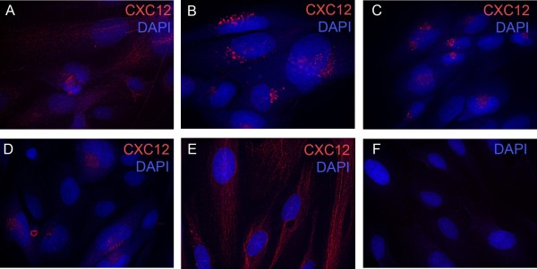
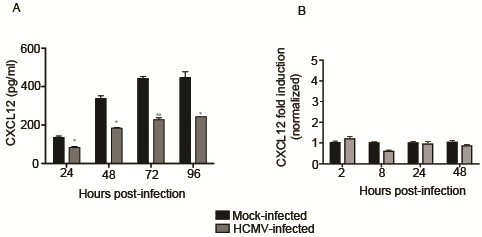
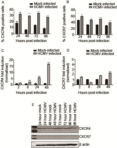
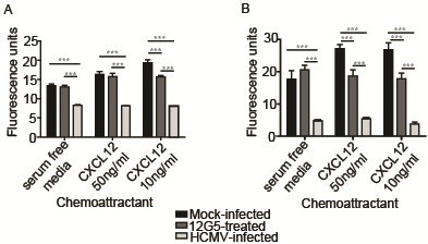
Results: Infection with HCMV significantly decreased secretion of CXCL12 by SGHPL-4 cells, and induced a striking perinuclear accumulation of the chemokine. HCMV infection significantly increased mRNA and total cell surface expression of the two known receptors for CXCL12: CXCR4 and CXCR7. Functionally, HCMV-infected SGHPL-4 cells were unable to migrate or invade in response to a gradient of soluble CXCL12 in transwell assays.

Conclusions: Collectively, these studies demonstrate that HCMV impairs EVT migration and invasion induced by CXCL12. As HCMV has the ability to inhibit EVT migration and invasion through dysregulation of other relevant signaling pathways, it is likely that the virus affects multiple signaling pathways to impair placentation and contribute to some of the placental defects seen in HCMV-positive pregnancies.


Figures

Figure 1
Figure 1
SGHPL-4 cells express CXCL12 and HCMV infection induces perinuclear sequestration of CXCL12 in SGHPL-4 cells. SGHPL-4 cells demonstrated positive staining for CXCL12 and mock-infected cells displayed diffuse staining for CXCL12 (A). SGHPL-4 cells were seeded on chamber slides and serum starved for 24 hours. They were infected with either laboratory (Towne and Toledo) or clinical (TRpM1a) isolates of HCMV for 24 hours, and then fixed and stained for CXCL12. In contrast, Towne (B), Toledo (C), and TRpMIA (D) were able to induce perinuclear sequestration of CXCL12. UV-inactivated Towne-GFP (E) was unable to induce chemokine sequestration. No significant background staining was seen in cells stained with secondary antibody and DAPI alone (F).
Figure 2
Figure 2
HCMV infection decreases secretion of CXCL12 by SGHPL-4 cells but mRNA expression is unaffected. Supernatants were collected from mock-infected or HCMV-infected cells and CXCL12 secretion was analyzed by ELISA (A). At all timepoints studied, HCMV-infected cells secreted significantly less CXCL12 into their culture medium as compared to mock-infected controls. RNA was isolated from mock-infected and HCMV-infected cells and quantitative real-time PCR was performed using primers specific for CXCL12 (B). There was no significant difference in mRNA expression between mock-infected and HCMV-infected cells at any timepoint analyzed. (*, p<0.05; **, p<0.01; ***, p<0.001).
Figure 3
Figure 3
HCMV infection alters expression of CXCR4 and CXCR7. SGHPL-4 cells were mock-infected or infected with HCMV, and then CXCR4 and CXCR7 receptor expression was determined. HCMV infection induced upregulation of cell surface staining for CXCR4 (A) and CXCR7 (B) at all time points studied. (*, p<0.05; **, p<0.01; ***, p<0.001). Total RNA was harvested from mock-infected or HCMV-infected SGHPL-4 cells, and analyzed by real-time PCR using primers specific for CXCR4 and CXCR7. HCMV infection induced a significant upregulation of CXCR4 mRNA expression (C) at 8, 24, and 48 hours post-infection. A modest but significant upregulation of CXCR7 mRNA expression was seen at 48 hours post-infection (D). Protein lysates from mock-infected and HCMV-infected cells were analyzed by Western blot for CXCR4 and CXCR7 expression. No differences in absolute protein levels were seen at any time point analyzed (E).
Figure 4
Figure 4
HCMV infection blocks the ability of SGHPL-4 cells to migrate or invade toward CXCL12. Serum-starved SGHPL-4 cells were mock-infected or infected with Towne HCMV (MOI of 1) for 24 hours. Mock-infected cells were also pretreated with a neutralizing antibody against CXCR4 (12G5) prior to the assay. The cells were loaded into Fluoroblok inserts with 8 μm pores and chemoattractants (CXCL12 or EGF) were loaded into the lower chamber. Migration was allowed to proceed for 6 hours, whereas invasion assays were incubated for 24 hours, at which point they were labeled with Calcein AM and 10X images were acquired. HCMV infection significantly impaired migration (A) and invasion (B) toward CXCL12 at both 50 ng/ml and 10 ng/ml (p<0.0001). Although antibody-mediated (12G5) neutralization of CXCR4 significantly decreased migration (p<0.0001) and invasion (p<0.0001) toward CXCL12, its effect was not as pronounced as that seen following infection with HCMV. (***, p<0.001).

References

    1. Demmler GJ. Congenital cytomegalovirus infection and disease. Adv Pediatr Infect Dis. 1996;11:135–162. - PubMed
    1. Gabrielli L, Bonasoni MP, Lazzarotto T, Lega S, Santini D, Foschini MP, Guerra B, Baccolini F, Piccirilli G, Chiereghin A. et al. Histological findings in foetuses congenitally infected by cytomegalovirus. J Clin Virol. 2009;46(Suppl 4):S16–S21. - PubMed
    1. von Dadelszen P, Magee LA. Could an infectious trigger explain the differential maternal response to the shared placental pathology of preeclampsia and normotensive intrauterine growth restriction? Acta Obstet Gynecol Scand. 2002;81:642–648. - PubMed
    1. Staun-Ram E, Shalev E. Human trophoblast function during the implantation process. Reprod Biol Endocrinol. 2005;3:56. doi: 10.1186/1477-7827-3-56. - DOI - PMC - PubMed
    1. Genbacev O, Miller RK. Post-implantation differentiation and proliferation of cytotrophoblast cells: in vitro models a review. Placenta. 2000;21(Suppl A):S45–S49. - PubMed

Publication types