. 2012 Nov 1:13:137.
doi: 10.1186/1471-2202-13-137.

A novel rodent model of spinal metastasis and spinal cord compression

Affiliations

A novel rodent model of spinal metastasis and spinal cord compression

Zion Zibly et al. BMC Neurosci. .

Abstract

Background: Spinal cord metastatic lesions affect a high number of cancer patients usually resulting in spinal cord compression syndrome. A major obstacle in the research of spinal metastatic disease is the lack of a simple reproducible animal model that mimics the natural course of the disease. In this study, we present a highly reproducible rodent model that can be used for different types of cancers while mimicking the natural course of human metastatic spinal cord compression syndrome.

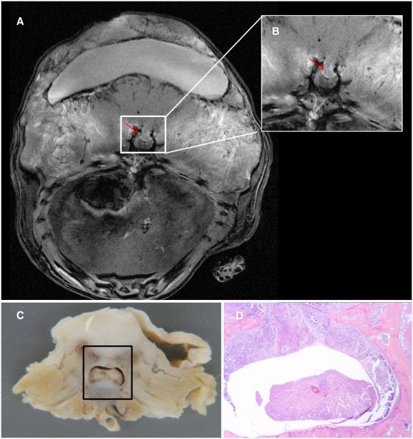
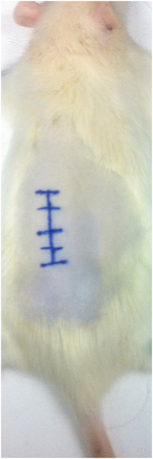
Results: All sixteen Fisher 344 rats survived the dorsal approach intraosseous implantation of CRL-1666 adenocarcinoma cells and both rats survived the sham control surgery. By Day 13 functional analysis via the modified Basso-Beattie-Bresnahan (BBB) locomotor rating scale showed significant decrease in motor function; median functional score was 3 for the tumor group (p = 0.0011). Median time to paresis was 8.7 days post-operatively. MR imaging illustrated repeated and consistent tumor formation, furthermore, onset of neurological sequale was the result of tumor formation and cord compression as confirmed by histological examination.

Conclusions: Analysis of these findings demonstrates a repeatable and consistent tumor growth model for cancer spinal metastases in rats. This novel rat model requires a less intricate surgical procedure, and as a result minimizes procedure time while subsequently increasing consistency. Therefore, this model allows for the preclinical evaluation of therapeutics for spinal metastases that more closely replicates physiological findings.


Figures

Figure 1
Figure 1
The median BBB score of treatment group over time from date of surgery until MR imaging. Cut-off value was a BBB score of 3.
Figure 2
Figure 2
T1 weighted post contrast MRI showing the destruction of the inner cortex of the laminae and cord compression by the tumor (red arrow in both frames). (A) Higher magnification of the spinal cord and invading tumor compressing the cord (B) Gross anatomical section of rat spinal cord showing tumor invasion and destruction of the vertebra as well as spinal cord compression (outlined box). (C) Histological cross-section of rat vertebra exhibiting tumor infiltration (T; stained purple) into the spinal canal and vertebra (pink) (D).
Figure 3
Figure 3
Animal positioning and preparation prior to surgical procedure. The thoraco-lumbar back is shaved, prepared and the mid-line skin above the spinal process is marked.
Figure 4
Figure 4
Skin incision above the spinal process and exposing the lamina of the vertebra bilateral. (A) Preparing the lamina for tumor transplant, drilling of the right lamina outer cortex after resection of the spinous process. (B).
Figure 5
Figure 5
Artist rendering of surgical procedure. (A) Drilling of the outer cortex of the lamina. (B) Implantation of the tumor cells and covering by bone wax. (C) Tumor growth and spinal cord pressure.

References

    1. Coleman RE. Metastatic bone disease: clinical features, pathophysiology and treatment strategies. Cancer Treat Rev. 2001;27(3):165–176. doi: 10.1053/ctrv.2000.0210. - DOI - PubMed
    1. Mundy GR. Metastasis to bone: causes, consequences and therapeutic opportunities. Nat Rev Cancer. 2002;2(8):584–593. doi: 10.1038/nrc867. - DOI - PubMed
    1. Ushio Y, Posner R, Kim JH, Shapiro WR, Posner JB. Treatment of experimental spinal cord compression caused by extradural neoplasms. J Neurosurg. 1977;47(3):380–390. doi: 10.3171/jns.1977.47.3.0380. - DOI - PubMed
    1. Ushio Y, Posner R, Posner JB, Shapiro WR. Experimental spinal cord compression by epidural neoplasm. Neurology. 1977;27(5):422–429. doi: 10.1212/WNL.27.5.422. - DOI - PubMed
    1. Mantha A, Legnani FG, Bagley CA, Gallia GL, Garonzik I, Pradilla G, Amundson E, Tyler BM, Brem H, Gokaslan ZL. A novel rat model for the study of intraosseous metastatic spine cancer. J Neurosurg Spine. 2005;2(3):303–307. doi: 10.3171/spi.2005.2.3.0303. - DOI - PubMed

Publication types

LinkOut - more resources