. 2012 Nov 2:8:81.
doi: 10.1186/1744-8069-8-81.

Cutaneous nociceptors lack sensitisation, but reveal μ-opioid receptor-mediated reduction in excitability to mechanical stimulation in neuropathy

Affiliations

Cutaneous nociceptors lack sensitisation, but reveal μ-opioid receptor-mediated reduction in excitability to mechanical stimulation in neuropathy

Yvonne Schmidt et al. Mol Pain. .

Abstract

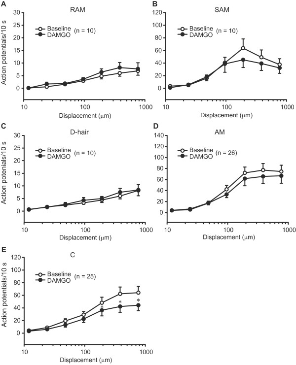
Background: Peripheral nerve injuries often trigger a hypersensitivity to tactile stimulation. Behavioural studies demonstrated efficient and side effect-free analgesia mediated by opioid receptors on peripheral sensory neurons. However, mechanistic approaches addressing such opioid properties in painful neuropathies are lacking. Here we investigated whether opioids can directly inhibit primary afferent neuron transmission of mechanical stimuli in neuropathy. We analysed the mechanical thresholds, the firing rates and response latencies of sensory fibres to mechanical stimulation of their cutaneous receptive fields.

Results: Two weeks following a chronic constriction injury of the saphenous nerve, mice developed a profound mechanical hypersensitivity in the paw innervated by the damaged nerve. Using an in vitro skin-nerve preparation we found no changes in the mechanical thresholds and latencies of sensory fibres from injured nerves. The firing rates to mechanical stimulation were unchanged or reduced following injury. Importantly, μ-opioid receptor agonist [D-Ala2,N-Me-Phe4,Gly5]-ol-enkephalin (DAMGO) significantly elevated the mechanical thresholds of nociceptive Aδ and C fibres. Furthermore, DAMGO substantially diminished the mechanically evoked discharges of C nociceptors in injured nerves. These effects were blocked by DAMGO washout and pre-treatment with the selective μ-opioid receptor antagonist Cys2-Tyr3-Orn5-Pen7-amide. DAMGO did not alter the responses of sensory fibres in uninjured nerves.

Conclusions: Our findings suggest that behaviourally manifested neuropathy-induced mechanosensitivity does not require a sensitised state of cutaneous nociceptors in damaged nerves. Yet, nerve injury renders nociceptors sensitive to opioids. Prevention of action potential generation or propagation in nociceptors might represent a cellular mechanism underlying peripheral opioid-mediated alleviation of mechanical hypersensitivity in neuropathy.


Figures

Figure 1
Figure 1
In vivo mechanical hypersensitivity following nerve injury. Measurements were performed with von Frey hairs applied to the plantar (A) and dorsal (B) surfaces of hind paws, before and 2 weeks after CCI or sham operation on the saphenous nerve. *P < 0.05, compared to thresholds before injury, thresholds of contralateral paws of CCI animals, and of both hind paws of sham-operated animals (2-way RM ANOVA, Bonferroni t test). Data are expressed as means ± SEM. N = 6 mice per group.
Figure 2
Figure 2
Conduction velocity and mechanical thresholds of sensory fibres following nerve injury. (A) Conduction velocities of Aβ and Aδ, but not of C fibres, were slightly decreased in injured nerves (*P < 0.05 and P > 0.05, respectively; Mann–Whitney test). (B) Mechanical von Frey thresholds of sensory fibres were not altered by nerve injury (P > 0.05; Mann–Whitney test). In uninjured nerves, the number of fibres from sham-operated and nonoperated nerves is as follows: Aβ fibres (18 and 50), Aδ fibres (12 and 53), and C fibres (5 and 14). All data are expressed as means ± SEM. N, number of fibres.
Figure 3
Figure 3
Mechanical latency of sensory fibres following nerve injury. Latencies of RAM and SAM (Aβ), D-hair and AM (Aδ), and C fibres to nanomotor stimulation were not significantly altered by nerve injury (P > 0.05; 2-way RM ANOVA) (A-E). In uninjured nerves, the number of fibres from sham-operated and nonoperated nerves is as follows: RAM (4 and 14), SAM (12 and 11), D-hair (4 and 7), AM (8 and 18), and C fibres (4 and 7). Data are expressed as means ± SEM. N, number of fibres.
Figure 4
Figure 4
Discharges of sensory fibres to mechanical stimulation following nerve injury. Discharge rates to nanomotor stimulation were not significantly altered in RAM (Aβ), D-hair (Aδ) and C fibres (P > 0.05; 2-way RM ANOVA) (A, C, E), but were diminished in SAM (Aβ) and AM (Aδ) fibres in injured nerves (*P < 0.05, compared to uninjured nerves; 2-way RM ANOVA, Bonferroni t test) (B, D). In uninjured nerves, the number of fibres from sham-operated and nonoperated nerves is the same as in Figure 3. All data are expressed as means ± SEM. N, number of fibres.
Figure 5
Figure 5
Effects of DAMGO on mechanical thresholds of sensory fibres following nerve injury. (A) Percentages of DAMGO-responding fibres in uninjured and injured nerves. The number of Aδ and C, but not of Aβ, fibres in which DAMGO (100 μM) increased von Frey thresholds was significantly higher in injured compared to uninjured nerves (P < 0.05; Fisher exact test for C fibres, and chi-square test for Aβ and Aδ fibres; calculated on raw data). In uninjured nerves, the number of fibres from sham-operated and nonoperated nerves is the same as in Figure 2. (B) Elevation of mechanical thresholds of Aδ and C fibres in injured nerves by DAMGO (100 μM), and its blockade by DAMGO washout or pre-treatment with μ-opioid receptor antagonist CTOP (100 μM). All Aδ fibres were classified as AM nociceptors. *P < 0.05, compared to all other conditions (1-way RM ANOVA, Bonferroni t test for Aδ fibres, and 1-way RM ANOVA on ranks, Tukey test for C fibres). Data are expressed as means ± SEM. N, number of fibres.
Figure 6
Figure 6
Effects of DAMGO on mechanical latencies of sensory fibres in injured nerves. Baseline mechanical latencies of Aβ (RAM, SAM), Aδ (D-hair, AM) and C fibres to nanomotor stimulation were not significantly changed by DAMGO (100 μM) (P > 0.05; 2-way RM ANOVA) (A-E). Data are expressed as means ± SEM. N, number of fibres.
Figure 7
Figure 7
Effects of DAMGO on discharges of sensory fibres in injured nerves. Baseline discharge rates of Aβ (RAM, SAM) and Aδ (D-hair, AM) fibres to nanomotor stimulation were not significantly altered by DAMGO (100 μM) (P > 0.05; 2-way RM ANOVA) (A-D). DAMGO significantly diminished the discharges of C fibres (*P < 0.05, 2-way RM ANOVA, Bonferroni t test) (E). All data are expressed as means ± SEM. N, number of fibres.
Figure 8
Figure 8
Effects of DAMGO on discharges of AM (Aδ) fibres with elevated mechanical thresholds in injured nerves. Baseline discharge rates of AM nociceptors to the nanomotor stimulation were not significantly changed by DAMGO (100 μM) or control buffer (P > 0.05; 2-way RM ANOVA). Data are expressed as means ± SEM. N, number of fibres.
Figure 9
Figure 9
Effects of DAMGO on discharges of C fibres with elevated mechanical thresholds in injured nerves. (A) Representative examples of C fibre firing (upper panels) and quantitative analysis (lower panels) showing that baseline discharge rates of C nociceptors to nanomotor stimulation were diminished by DAMGO (100 μM) (*P < 0.05; 2-way RM ANOVA, Bonferroni t test), but not by control buffer (P > 0.05; 2-way RM ANOVA). DAMGO and buffer were tested on separate sets of fibres. In upper panels the displacements are marked with dark lines, and the interstimulus sequences are removed. (B) DAMGO (100 μM)-induced reduction in the discharge rate was reversed by DAMGO washout and prevented by pre-application of CTOP (100 μM). This was tested in additional C fibres at a single nanomotor stimulation (384 μm). P < 0.05, compared to all other conditions (1-way RM ANOVA; Bonferroni t test). All data are expressed as means ± SEM. N, number of fibres.

References

    1. Ochoa JL, Yarnitsky D. Mechanical hyperalgesias in neuropathic pain patients: dynamic and static subtypes. Ann Neurol. 1993;33:465–472. doi: 10.1002/ana.410330509. - DOI - PubMed
    1. Baron R, Binder A, Wasner G. Neuropathic pain: diagnosis, pathophysiological mechanisms, and treatment. Lancet Neurol. 2010;9:807–819. doi: 10.1016/S1474-4422(10)70143-5. - DOI - PubMed
    1. Dowdall T, Robinson I, Meert TF. Comparison of five different rat models of peripheral nerve injury. Pharmacol Biochem Behav. 2005;80:93–108. doi: 10.1016/j.pbb.2004.10.016. - DOI - PubMed
    1. Koltzenburg M, Bennett DL, Shelton DL, McMahon SB. Neutralization of endogenous NGF prevents the sensitization of nociceptors supplying inflamed skin. Eur J Neurosci. 1999;11:1698–1704. doi: 10.1046/j.1460-9568.1999.00590.x. - DOI - PubMed
    1. LaMotte RH, Lundberg LE, Torebjork HE. Pain, hyperalgesia and activity in nociceptive C units in humans after intradermal injection of capsaicin. J Physiol. 1992;448:749–764. - PMC - PubMed

Publication types

Substances