

Discovery of SLC3A2 cell membrane protein as a potential gastric cancer biomarker: implications in molecular imaging
- PMID: 23116296
- PMCID: PMC3749780
- DOI: 10.1021/pr300555y
Discovery of SLC3A2 cell membrane protein as a potential gastric cancer biomarker: implications in molecular imaging
Abstract
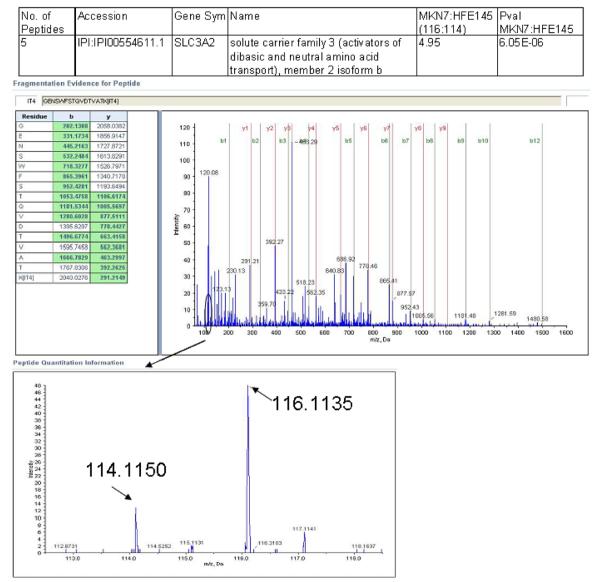
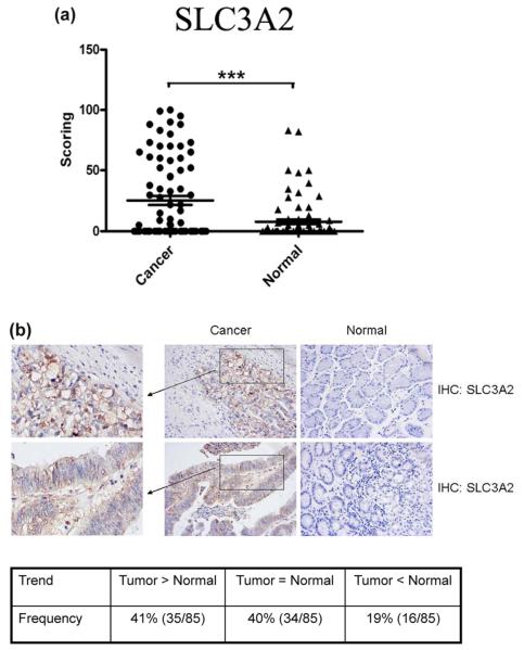
Despite decreasing incidence and mortality, gastric cancer remains the second leading cause of cancer-related deaths in the world. Successful management of gastric cancer is hampered by lack of highly sensitive and specific biomarkers especially for early cancer detection. Cell surface proteins that are aberrantly expressed between normal and cancer cells are potentially useful for cancer imaging and therapy due to easy accessibility of these targets. Combining two-phase partition and isobaric tags for relative and absolute quantification methods, we compared the relative expression levels of membrane proteins between noncancer and gastric cancer cells. About 33% of the data set was found to be plasma membrane and associated proteins using this approach (compared to only 11% in whole cell analysis), several of which have never been previously implicated in gastric cancer. Upregulation of SLC3A2 in gastric cancer cells was validated by immunoblotting of a panel of 13 gastric cancer cell lines and immunohistochemistry on tissue microarrays comprising 85 matched pairs of normal and tumor tissues. Immunofluorescence and immunohistochemistry both confirmed the plasma membrane localization of SLC3A2 in gastric cancer cells. The data supported the notion that SLC3A2 is a potential biomarker that could be exploited for molecular imaging-based detection of gastric cancer.
Figures


References
-
- Ferlay J, Shin HR, Bray F, Forman D, Mathers C, Parkin DM. Estimates of worldwide burden of cancer in 2008: GLOBOCAN 2008. Int. J. Cancer. 2010;127(12):2893–917. - PubMed
-
- Akdogan M, Sasmaz N, Kayhan B, Biyikoglu I, Disibeyaz S, Sahin B. Extraordinarily elevated CA19-9 in benign conditions: a case report and review of the literature. Tumori. 2001;87(5):337–339. - PubMed
-
- Kim JM, Sohn HY, Yoon SY, Oh JH, Yang JO, Kim JH, Song KS, Rho SM, Yoo HS, Kim YS, Kim JG, Kim NS. Identification of gastric cancer-related genes using a cDNA microarray containing novel expressed sequence tags expressed in gastric cancer cells. Clin. Cancer Res. 2005;11(2 Pt 1):473–482. - PubMed
-
- Tseng CW, Yang JC, Chen CN, Huang HC, Chuang KN, Lin CC, Lai HS, Lee PH, Chang KJ, Juan HF. Identification of 14-3-3beta in human gastric cancer cells and its potency as a diagnostic and prognostic biomarker. Proteomics. 2011;11(12):2423–2439. - PubMed
-
- Melle C, Ernst G, Schimmel B, Bleul A, Kaufmann R, Hommann M, Richter KK, Daffner W, Settmacher U, Claussen U, von Eggeling F. Characterization of pepsinogen C as a potential biomarker for gastric cancer using a histo-proteomic approach. J. Proteome Res. 2005;4(5):1799–1804. - PubMed
Publication types
MeSH terms
Substances
Grants and funding
LinkOut - more resources
Full Text Sources
Medical
Molecular Biology Databases
Research Materials
