Review
. 2013;19(14):2606-14.
doi: 10.2174/1381612811319140010.

Chemical nature and reaction mechanisms of the molybdenum cofactor of xanthine oxidoreductase

Affiliations
Free PMC article
Review

Chemical nature and reaction mechanisms of the molybdenum cofactor of xanthine oxidoreductase

Ken Okamoto et al. Curr Pharm Des. 2013.
Free PMC article

Abstract

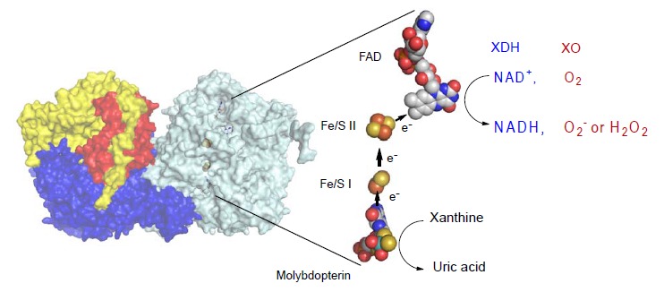
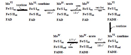
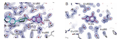
Xanthine oxidoreductase (XOR), a complex flavoprotein, catalyzes the metabolic reactions leading from hypoxanthine to xanthine and from xanthine to urate, and both reactions take place at the molybdenum cofactor. The enzyme is a target of drugs for therapy of gout or hyperuricemia. We review the chemical nature and reaction mechanisms of the molybdenum cofactor of XOR, focusing on molybdenum-dependent reactions of actual or potential medical importance, including nitric oxide (NO) synthesis. It is now generally accepted that XOR transfers the water-exchangeable -OH ligand of the molybdenum atom to the substrate. The hydroxyl group at OH-Mo(IV) can be replaced by urate, oxipurinol and FYX-051 derivatives and the structures of these complexes have been determined by xray crystallography under anaerobic conditions. Although formation of NO from nitrite or formation of xanthine from urate by XOR ischemically feasible, it is not yet clear whether these reactions have any physiological significance since the reactions are catalyzed at a slow rate even under anaerobic conditions.


Figures

Fig. (1)
Fig. (1)
Crystal Structure of Bovine XOR. Left; Homodimer structure of bovine XOR. The N-terminal (in red), the C-terminal (in blue) and the intermediate (in yellow) domains contain the iron-sulfur centers, the molybdopterin and the FAD centers. Right: Cofactor arrangements of the enzyme. Figures were generated from PDB ID 1F4Q. Arrows show the directions of electron flow during catalysis. The reduced FAD reacts with either NAD+ or oxygen to produce NADH or hydrogen peroxide (H2O2) or superoxide (O2-). FADH2 reacts with O2 to produce H2O2 , while FADH produces O2- [61, 63]. (The color version of the figure is available in the electronic copy of the article).
Fig. (2)
Fig. (2)
Hydroxylation Reaction of Purines. A; Hydroxylation order in purine catabolism. Accumulation of xanthine was observed, indicating that the reaction does not occur sequentially. The absence of formation of 6, 8-dihydroxypurine suggests that hydroxylation at the 2-position is important for hydroxylation at the 8-position. B; Concentrations of hypoxanthine, xanthine and urate were measured with HPLC during turnover from hypoxanthine to urate catalyzed by bovine milk xanthine oxidase. (T. Kusano, unpublished results).
Fig. (3)
Fig. (3)
Schematic Diagram of the Reaction of Molybdenum during Xanthine Hydroxylation. The results of fast reaction between xanthine and chicken xanthine dehydrogenase followed by means of a stopped-flow method at 4°C. Dissociation of urate (1.8/sec) is the slowest step, and is rate-limiting for the overall process. This figure is drawn based on reference [29].
Fig. (4)
Fig. (4)
Structure of Mammalian Molybdopterin and Hydroxylation Mechanism of Artificial Slow Substrate FYX-051. A; Chemical structure of mammalian XOR molybdopterin. B; Geometry of molybdenum-coodinated atoms in the oxidized state (left) and reduced state (right). C; Glu1261 works as a base, abstracting the proton from Mo-OH (a). The generated Mo-O- nucleophilically attacks the carbon center to be hydroxylated, with concomitant hydride transfer (b). The protonated Glu1261 forms a hydrogen bond to the N1-nitrogen of the substrate, and this facilitates the nucleophilic attack (b). The reduced Mo(IV) coordinated to the product via the newly introduced hydroxyl group (c). The intermediate breaks down by hydroxide displacement of the product (d).
Fig. (5)
Fig. (5)
Two Hydroxylation Models of Xanthine Hydroxylation. A; Proposed model of xanthine binding mode based on the analysis of mutant enzymes, as well as the urate binding mode. The hydrogen bonds of the three amino acids promote nucleophilic reaction at C8 (based on 38, 45). B; Activation of substrate xanthine by Arg881 via accumulation of negative charge at the 6-position oxygen (based on 39). C; Proposed hypoxanthine binding mode based on the analysis of mutant enzymes (based on 38) and binding mode of hypoxanthine to the desulfo-form in the crystal (Fig. 5E). D; Activation of substrate hypoxanthine owing to accumulation of negative charge at the 2-position oxygen. The crystal structure of a different binding mode from C was also reported (based on 42). E; Crystal structure of hypoxanthine bound bovine desulfo-XOR, which lacks an essential sulfur atom at the active site, at 2.0 Å resolution (unpublished data). The 2Fo-Fc electron density map was contoured at 1.3 σ. A hydrogen bond is shown as a broken line.
Fig. (6)
Fig. (6)
Proposed Hydrogen-Bonding Arrangement of the Xanthine-bound Complex with Molybdopterin, and Mechanism of the Xanthine Hydroxylation Based on this Binding Mode. Glu1261 abstracts the proton from Mo-OH (a). The -O- thus generated is then involved in electrophilic attack on the C8 carbon of xanthine with hydride transfer to the =S of the molybdopterin (b), resulting in a covalent linkage between the molybdenum ion and the C8 carbon atom via the bridging oxygen atom (c). The protonated Glu1261 and glutamate Glu802, which is also supposed to be protonated under physiological conditions, form hydrogen bonds to the substrate, stabilizing this state. Arg880, too, is involved in the hydroxylation by forming a hydrogen bond with the O2 atom of the xanthine molecule. The intermediate decomposes via the replacement of the bridge oxygen with a water molecule (d or e~ f).
Fig. (7)
Fig. (7)
Binding Modes of Urate with the Demolybdo Form of the Enzyme. A; The structure of the complex of urate bound to the reduced bovine XOR under anaerobic conditions was also determined. The 2Fo-Fc electron density map contoured at 1.0 σ. B; As urate dissociates from the holoenzyme without forming a stable binding mode, the X-ray crystal structure of the urate-bound form of rat XOR D428A mutant enzyme without the molybdenum cofactor (demolybdo enzyme) was determined. The 2Fo-Fc electron density map was contoured at 1.5 σ. Figures were generated from PDB ID 3AMZ and 3AN1, respectively (45).
Fig. (8)
Fig. (8)
Summary of the Chemical Structures of Ligands Covalently Bound to Reduced Molybdenum in the Active Site of XOR. A; urate, B: oxipurinol, C: FYX-051, D: trihydroxy-FYX-051. E; proposed mechanism of NO formation by XO based on reference [57]. Related crystal structures are shown below. Figures were generated from PDB ID 3AMZ, 3BDJ, 1V97 and 3AM9, respectively (45, 51, 19, 52).

References

    1. Elion GB. The purine path to chemotherapy. Science. 1989;244:41–7. - PubMed
    1. Hille R. The mononuclear molybdenum enzymes. Chem Rev. 1996;96:2757–816. - PubMed
    1. Enroth C, Eger BT, Okamoto K, Nishino T, Nishino T, Pai EF. Structures of bovine milk xanthine dehydrogenase and xanthine oxidase structure based mechanism of conversion. Proc Natl Acad Sci USA. 2000;97:10723–8. - PMC - PubMed
    1. Porras AG, Palmer G. The room temperature potentiometry of xanthine oxidase: pH-dependent redox behavior of the flavin, molybdenum, and iron-sulfur centres. J Biol Chem. 1982;257:11617–26. - PubMed
    1. Hunt J, Massey V, Dunham WR, Sands RH. Redox potentials of milk xanthine dehydrogenase room temperature measurement of the FAD and 2 Fe/2 S center potentials. J Biol Chem. 1993;268:18685–91. - PubMed

Publication types

MeSH terms

LinkOut - more resources