

Case Reports
doi: 10.1093/icvts/ivs430.
Epub 2012 Nov 1.
Wavering calcified amorphous tumour of the heart in a haemodialysis patient
Affiliations
- PMID: 23117236
- PMCID: PMC3548521
- DOI: 10.1093/icvts/ivs430
Item in Clipboard
Case Reports
Wavering calcified amorphous tumour of the heart in a haemodialysis patient
Interact Cardiovasc Thorac Surg.
2013 Feb.
Abstract
Calcified amorphous tumour is a rare, non-neoplastic, endocardially based, intracavitary cardiac mass. This report describes a 59-year old man in whom a mobile mass was found incidentally in the heart by routine echocardiography after he had been on haemodialysis for 3 years. Transoesophageal echocardiography revealed a high-echoic swinging tumour that originated from the annulus of the anterior commissure of the mitral valve. Surgical resection was performed to prevent embolization, and his clinical course was excellent.
Figures
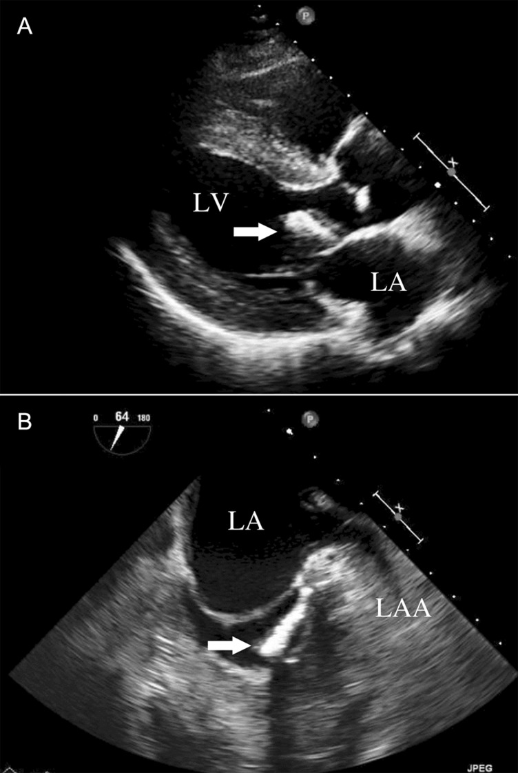
(A) Transthoracic echocardiogram. The long-axis view shows high-echoic mass (arrow) adhering to the mitral annulus of the anterior leaflet. (B) Transoesophageal echocardiogram. The short-axis view shows a cudgel-shaped, homogenous, high-echoic mass (arrow) that originated from the annulus of the anterior commissure of the mitral valve near the left fibrous trigone. Mitral annulus calcification was also recognized in the same area. LA: left atrium; LV: left ventricle; LAA: left atrial appendage.
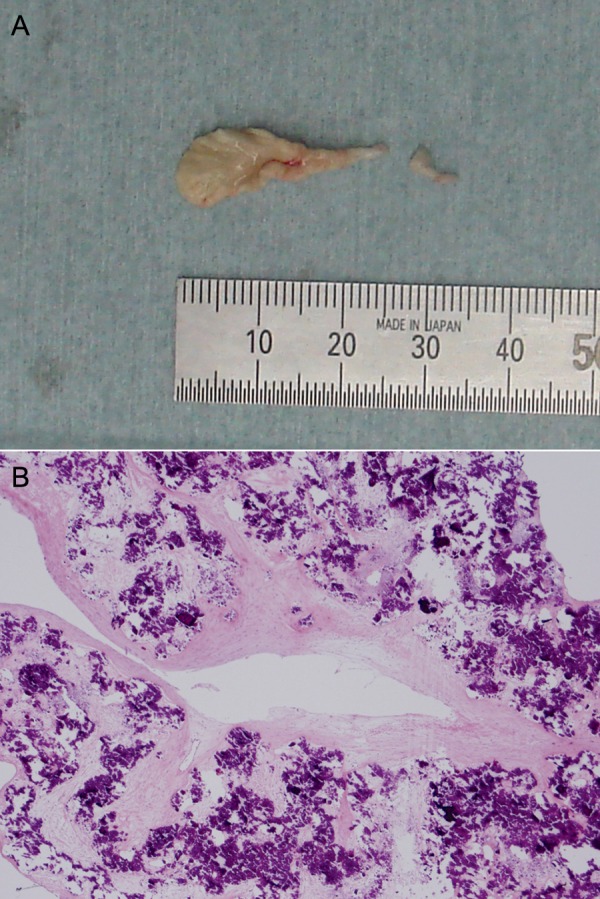
(A) Gross appearance of the resected tumour. (B) Histopathological appearance of the tumour. Fine deposits of calcium surrounded by amorphous fibrin and collagenous fibres, and chronic inflammation are seen (haematoxylin and eosin staining, ×40).
References
-
- Reynolds C, Tazelaar HD, Edwards WD. Calcified amorphous tumor of the heart (cardiac CAT) Hum Pathol. 1997;28:601–6. - PubMed
-
- Habib A, Friedman PA, Cooper LT, Suleiman L, Asirvatham SJ. Cardiac calcified amorphous tumor in a patient presenting for ventricular tachycardia ablation: intracardiac echocardiogram diagnosis and management. J Interv Card Electrophysiol. 2010;29:175–8. - PubMed
-
- Ho HH, Min JK, Lin F, Wong SC, Bergman G. Calcified amorphous tumor of the heart. Circulation. 2008;117:e171–2. - PubMed
-
- Kubota H, Fujioka Y, Yoshino H, Koji H, Yoshihara K, Tonari K, et al. Cardiac swinging calcified amorphous tumors in end-stage renal failure patients. Ann Thorac Surg. 2010;90:1692–4. - PubMed
-
- Fujiwara M, Watanabe H, Iino T, Kobukai Y, Ishibashi K, Yamamoto H, et al. Two case of calcified amorphous tumor mimicking mitral valve vegetation. Circulation. 2012;125:e432–4. - PubMed
Publication types
MeSH terms
LinkOut - more resources
Full Text Sources
Medical
