

Amygdala volume changes in posttraumatic stress disorder in a large case-controlled veterans group
- PMID: 23117638
- PMCID: PMC3647246
- DOI: 10.1001/archgenpsychiatry.2012.50
Amygdala volume changes in posttraumatic stress disorder in a large case-controlled veterans group
Abstract
Context: Smaller hippocampal volumes are well established in posttraumatic stress disorder (PTSD), but the relatively few studies of amygdala volume in PTSD have produced equivocal results.
Objective: To assess a large cohort of recent military veterans with PTSD and trauma-exposed control subjects, with sufficient power to perform a definitive assessment of the effect of PTSD on volumetric changes in the amygdala and hippocampus and of the contribution of illness duration, trauma load, and depressive symptoms.
Design: Case-controlled design with structural magnetic resonance imaging and clinical diagnostic assessments. We controlled statistically for the important potential confounds of alcohol use, depression, and medication use.
Setting: Durham Veterans Affairs Medical Center, which is located in proximity to major military bases.
Patients: Ambulatory patients (n = 200) recruited from a registry of military service members and veterans serving after September 11, 2001, including a group with current PTSD (n = 99) and a trauma-exposed comparison group without PTSD (n = 101).
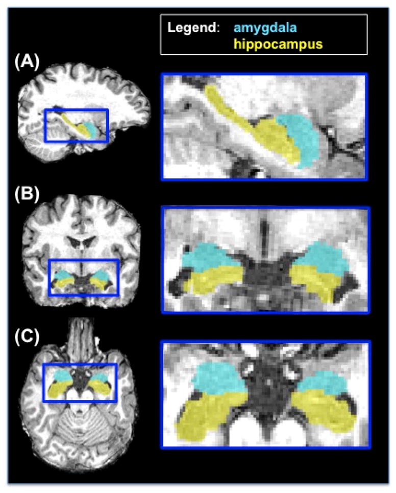
Main outcome measure: Amygdala and hippocampal volumes computed from automated segmentation of high-resolution structural 3-T magnetic resonance imaging.
Results: Smaller volume was demonstrated in the PTSD group compared with the non-PTSD group for the left amygdala (P = .002), right amygdala (P = .01), and left hippocampus (P = .02) but not for the right hippocampus (P = .25). Amygdala volumes were not associated with PTSD chronicity, trauma load, or severity of depressive symptoms.
Conclusions: These results provide clear evidence of an association between a smaller amygdala volume and PTSD. The lack of correlation between trauma load or illness chronicity and amygdala volume suggests that a smaller amygdala represents a vulnerability to developing PTSD or the lack of a dose-response relationship with amygdala volume. Our results may trigger a renewed impetus for investigating structural differences in the amygdala, its genetic determinants, its environmental modulators, and the possibility that it reflects an intrinsic vulnerability to PTSD.
Conflict of interest statement
The authors have no conflicts of interest and no financial disclosures. The entities that provided funding support had no input into the design and conduct of the study; collection, management, analysis, and interpretation of the data; and preparation, review, or approval of the manuscript. The principal investigator (Rajendra Morey, M.D.) had full access to all of the data in the study and takes responsibility for the integrity of the data and the accuracy of the data analysis.
Figures
References
-
- Rauch SL, Shin LM, Phelps EA. Neurocircuitry models of posttraumatic stress disorder and extinction: human neuroimaging research--past, present, and future. Biological Psychiatry. 2006 Aug 15;60(4):376–382. - PubMed
-
- LaBar KS, Gatenby JC, Gore JC, LeDoux JE, Phelps EA. Human amygdala activation during conditioned fear acquisition and extinction: a mixed-trial fMRI study. Neuron. 1998;20(5):937–945. - PubMed
-
- McEwen BS. Physiology and neurobiology of stress and adaptation: Central role of the brain. Physiol Rev. 2007 Jul;87(3):873–904. - PubMed
Publication types
MeSH terms
Grants and funding
LinkOut - more resources
Full Text Sources
Other Literature Sources
Medical
