. 2013 Feb;27(2):665-71.
doi: 10.1096/fj.12-220350. Epub 2012 Nov 1.

Adaptive radiation-induced epigenetic alterations mitigated by antioxidants

Affiliations

Adaptive radiation-induced epigenetic alterations mitigated by antioxidants

Autumn J Bernal et al. FASEB J. 2013 Feb.

Abstract

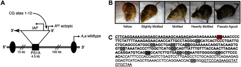
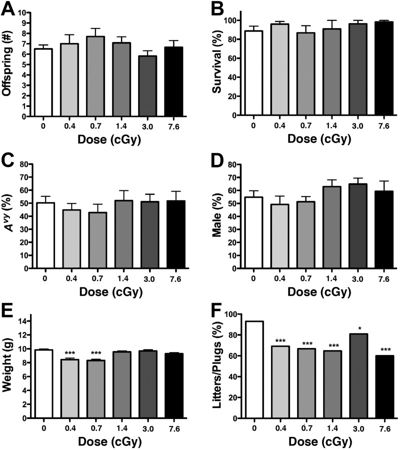
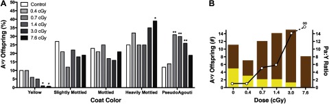
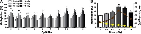
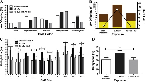
Humans are exposed to low-dose ionizing radiation (LDIR) from a number of environmental and medical sources. In addition to inducing genetic mutations, there is concern that LDIR may also alter the epigenome. Such heritable effects early in life can either be positively adaptive or result in the enhanced formation of diseases, including cancer, diabetes, and obesity. Herein, we show that LDIR significantly increased DNA methylation at the viable yellow agouti (A(vy)) locus in a sex-specific manner (P=0.004). Average DNA methylation was significantly increased in male offspring exposed to doses between 0.7 and 7.6 cGy, with maximum effects at 1.4 and 3.0 cGy (P<0.01). Offspring coat color was concomitantly shifted toward pseudoagouti (P<0.01). Maternal dietary antioxidant supplementation mitigated both the DNA methylation changes and coat color shift in the irradiated offspring. Thus, LDIR exposure during gestation elicits epigenetic alterations that lead to positive adaptive phenotypic changes that are negated with antioxidants, indicating they are mediated in part by oxidative stress. These findings provide evidence that in the isogenic A(vy) mouse model, epigenetic alterations resulting from LDIR play a role in radiation hormesis, bringing into question the assumption that every dose of radiation is harmful.


Figures

Figure 1.
Figure 1.
Avy locus and resulting mouse coat colors. A) Agouti gene encodes for a paracrine-signaling molecule that produces either black eumelanin (a) or yellow phaeomelanin (A) from the wild-type promoter (arrow labeled A,a wild type; refs. 9, 11). Agouti expression during follicle development results in brown (agouti) wild-type animals. Avy allele resulted from a spontaneous contraoriented insertion of an intracisternal A particle (IAP) into pseudoexon PS1A upstream of the wild-type promoter. This insertion carries a cryptic promoter (arrow labeled Avy ectopic) controlled by the methylation of upstream CpG sites. B) Level of CpG methylation at the Avy locus results in the formation of distinct coat color phenotypes. Hypermethylation of the cryptic IAP promoter results in brown, pseudoagouti offspring, hypomethylation results in yellow offspring, and mottled mice are epigenetic mosaic offspring. C) Amplified IAP sequence (bold font) contains 11 CpG sites in the ectopic promoter and 1 CpG site within the downstream 3′ genomic sequence (regular font). The first CpG site (highlighted in red) was not measured due to technical limitations associated with our sequencing approach. The eighth CpG site (highlighted in dark gray) was measured on the reverse strand.
Figure 2.
Figure 2.
Effect of radiation on litter size at weaning (A), percentage of offspring survival to weaning (B), percentage of offspring with an Avy genotype (C), percentage of male offspring (D), offspring weight at weaning (E), and percentage of plugged females with litters (F). *P < 0.05, ***P < 0.0001; χ2, ANOVA.
Figure 3.
Figure 3.
Effect of radiation on the coat color of Avy offspring. A) Percentage of yellow, slightly mottled, mottled, heavily mottled, and pseudoagouti Avy offspring at 0 cGy (n=47), 0.4 cGy (n=28), 0.7 cGy (n=32), 1.4 cGy (n=40), 3.0 cGy (n=53), and 7.6 cGy (n=41) was compared with that in the sham-irradiated offspring. *P < 0.05, **P < 0.01; χ2. B) Number of pseudoagouti (brown bars) and yellow (yellow bars) offspring at each radiation dose (left y axis) were used to calculate the pseudoagouti:yellow offspring ratio (open circles; right y axis) at each radiation dose.
Figure 4.
Figure 4.
Effect of radiation on DNA methylation and the coat color of Avy male offspring. A) Percentage methylation ± se of 11 CpG sites at the Avy locus in liver tissue from male offspring exposed to 0.4 cGy (n=13), 0.7 cGy (n=22), 1.4 cGy (n=24), 3.0 cGy (n=29), and 7.6 cGy (n=19) compared with male sham-irradiated offspring (n=26). *P ≤ 0.05, **P < 0.01. B) Average percentage methylation ± se of the 11 CpG sites in male liver tissue at various radiation doses was compared with that in sham-irradiated offspring (ANOVA, P<0.0001). Yellow and brown circles depict the percentage of the male Avy offspring with yellow and brown coat colors, respectively. *P < 0.05, **P < 0.01; Fisher's PLSD.
Figure 5.
Figure 5.
Effect of radiation and AO supplementation on DNA methylation and coat color of Avy offspring. A) Percentage of Avy offspring that were yellow, slightly mottled, mottled, heavily mottled, and pseudoagouti when the mothers were sham irradiated (n=53) and exposed to 3.0 cGy (n=80) or 3.0 cGy + AO (n=51). *P < 0.05, **P < 0.01; χ2. B) Number of pseudoagouti (brown bars) and yellow (yellow bars) offspring when the mothers were sham irradiated and exposed to 3·0 cGy or 3.0 cGy + AO (left y axis) were used to calculate the pseudoagouti:yellow offspring ratio (open circles; right y axis) for each exposure. C) Percentage methylation ± se of 11 CpG sites at the Avy locus in the liver tissue of male offspring sham-irradiated (n=27), and exposed to 3·0 cGy (n=38) or 3·0 cGy + AO (n=23). *P ≤ 0.05; ANOVA. D) Average percentage methylation ± se of 11 CpG sites at the Avy locus in the liver tissue of male offspring sham-irradiated (n=27), and exposed to 3.0 cGy (n=38) or 3.0 cGy + AO (n=23). *P ≤ 0.05; ANOVA.

References

    1. Schauer D. A., Linton O. W. (2009) NCRP report no. 160, ionizing radiation exposure of the population of the United States, medical exposure–are we doing less with more, and is there a role for health physicists? Health Phys. 97, 1–5 - PubMed
    1. Saenko V., Ivanov V., Tsyb A., Bogdanova T., Tronko M., Demidchik Y., Yamashita S. (2011) The Chernobyl accident and its consequences. Clin. Oncol. (R. Coll. Radiol.) 23, 234–243 - PubMed
    1. Akiba S. (2012) Epidemiological studies of Fukushima residents exposed to ionising radiation from the Fukushima Daiichi nuclear power plant prefecture–a preliminary review of current plans. J. Radiol. Prot. 32, 1–10 - PubMed
    1. Sanders C. (2009) Radiation Hormesis and the Linear-No-Threshold Assumption, Springer, New York
    1. Tawa R., Kimura Y., Komura J., Miyamura Y., Kurishita A., Sasaki M. S., Sakurai H., Ono T. (1998) Effects of X-ray irradiation on genomic DNA methylation levels in mouse tissues. J. Radiat. Res. 39, 271–278 - PubMed

Publication types