MRI of the penis
- PMID: 23118102
- PMCID: PMC3746407
- DOI: 10.1259/bjr/63301362
MRI of the penis
Abstract
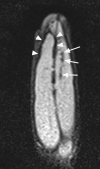
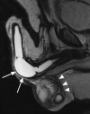
MRI of the penis is an expensive test that is not always superior to clinical examination or ultrasound. However, it shows many of the important structures, and in particular the combination of tumescence from intracavernosal alprostadil, and high-resolution T(2) sequences show the glans, corpora and the tunica albuginea well. In this paper we summarise the radiological anatomy and discuss the indications for MRI. For penile cancer, it may be useful in cases where the local stage is not apparent clinically. In priapism, it is an emerging technique for assessing corporal viability, and in fracture it can in most cases make the diagnosis and locate the injury. In some cases of penile fibrosis and Peyronie's disease, it may aid surgical planning, and in complex pelvic fracture may replace or augment conventional urethrography. It is an excellent investigation for the malfunctioning penile prosthesis.
Figures


Similar articles
-
MR imaging of nonmalignant penile lesions.Radiographics. 2008 May-Jun;28(3):837-53. doi: 10.1148/rg.283075100. Radiographics. 2008. PMID: 18480487 Review.
-
MRI of the Penis: Indications, Anatomy, and Pathology.Curr Probl Diagn Radiol. 2020 Jan-Feb;49(1):54-63. doi: 10.1067/j.cpradiol.2018.12.004. Epub 2018 Dec 11. Curr Probl Diagn Radiol. 2020. PMID: 30704768 Review.
-
[The role of magnetic resonance in the evaluation of pathological processes involving the penis].Clin Ter. 1993 Aug;143(2):167-71. Clin Ter. 1993. PMID: 8222546 Italian.
-
MR imaging of penile pathology and prostheses.Abdom Radiol (NY). 2025 Jan;50(1):305-318. doi: 10.1007/s00261-024-04417-2. Epub 2024 Jul 27. Abdom Radiol (NY). 2025. PMID: 39066812 Review.
-
MRI of the penis.Abdom Radiol (NY). 2020 Jul;45(7):2001-2017. doi: 10.1007/s00261-019-02301-y. Abdom Radiol (NY). 2020. PMID: 31701192 Review.
Cited by
-
Penile prosthesis surgery in the management of erectile dysfunction.Arab J Urol. 2013 Sep;11(3):245-53. doi: 10.1016/j.aju.2013.05.002. Epub 2013 Jul 2. Arab J Urol. 2013. PMID: 26558089 Free PMC article. Review.
-
What Is New in the Diagnosis and Management of Penile Cancer?Indian J Surg Oncol. 2017 Sep;8(3):379-384. doi: 10.1007/s13193-016-0613-2. Epub 2017 Feb 27. Indian J Surg Oncol. 2017. PMID: 36118381 Free PMC article. Review.
-
Partial thrombosis of the corpus cavernosum - a malignancy mimicker.BJR Case Rep. 2021 Sep 10;8(1):20210085. doi: 10.1259/bjrcr.20210085. eCollection 2022 Jan 1. BJR Case Rep. 2021. PMID: 35136638 Free PMC article.
-
Penile gangrene: an unusual complication of malignant priapism in a patient with renal cell carcinoma.Pan Afr Med J. 2019 Nov 5;34:130. doi: 10.11604/pamj.2019.34.130.20447. eCollection 2019. Pan Afr Med J. 2019. PMID: 33708299 Free PMC article.
-
Acute scrotal pain and priapism: an early sign of progression in metastatic renal cell carcinoma?BMJ Case Rep. 2015 Feb 18;2015:bcr2014208228. doi: 10.1136/bcr-2014-208228. BMJ Case Rep. 2015. PMID: 25694639 Free PMC article.
References
-
- Pretorius ES, Siegelman ES, Ramchandani P, Banner MP. MR imaging of the penis. Radiographics 2001;1:S283–98; discussion S98–9 - PubMed
-
- Velcek D, Evans JA. Cavernosography. Radiology 1982;144:781–5 - PubMed
-
- Kirkham AP, Illing R, Minhas S, Minhas S, Allen C. MR imaging of nonmalignant penile lesions. Radiographics 2008;28:837–53 - PubMed
-
- Horenblas S, Kroger R, Gallee MP, Newling DW, van Tinteren H. Ultrasound in squamous cell carcinoma of the penis; a useful addition to clinical staging? A comparison of ultrasound with histopathology. Urology 1994;43:702–7 - PubMed
-
- Bookstein JJ, Lang EV. Penile magnification pharmacoarteriography: details of intrapenile arterial anatomy. AJR Am J Roentgenol 1987;148:883–8 - PubMed
MeSH terms
LinkOut - more resources
Full Text Sources
Medical

