. 2013 Jan;12(1):228-36.
doi: 10.1074/mcp.M112.020651. Epub 2012 Nov 1.

Proteomic analysis of protease resistant proteins in the diabetic rat kidney

Affiliations

Proteomic analysis of protease resistant proteins in the diabetic rat kidney

Sneha B Bansode et al. Mol Cell Proteomics. 2013 Jan.

Abstract

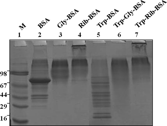
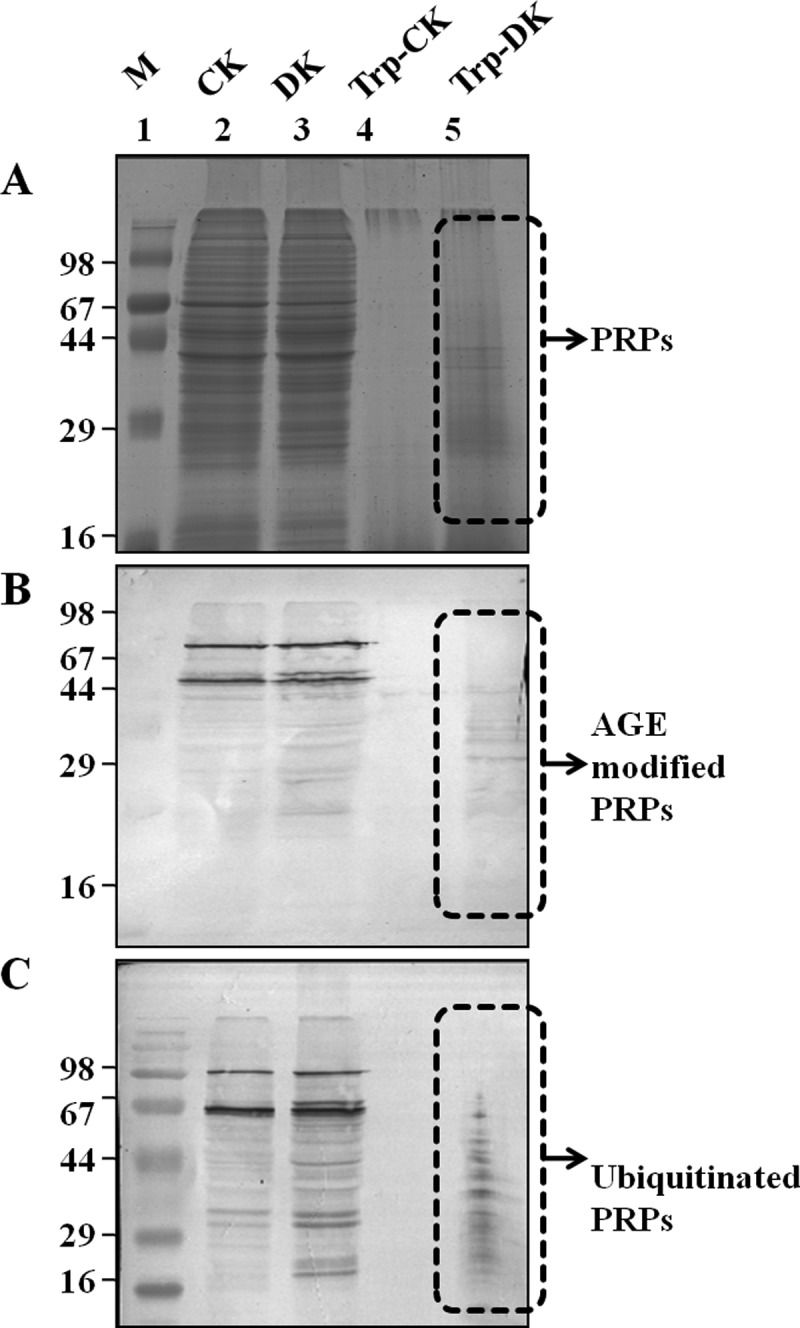
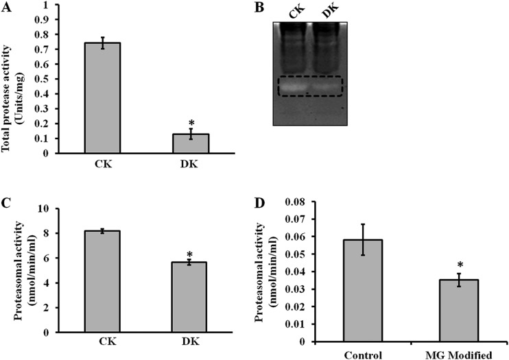
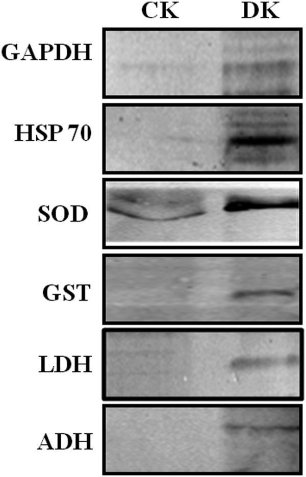
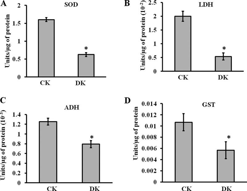
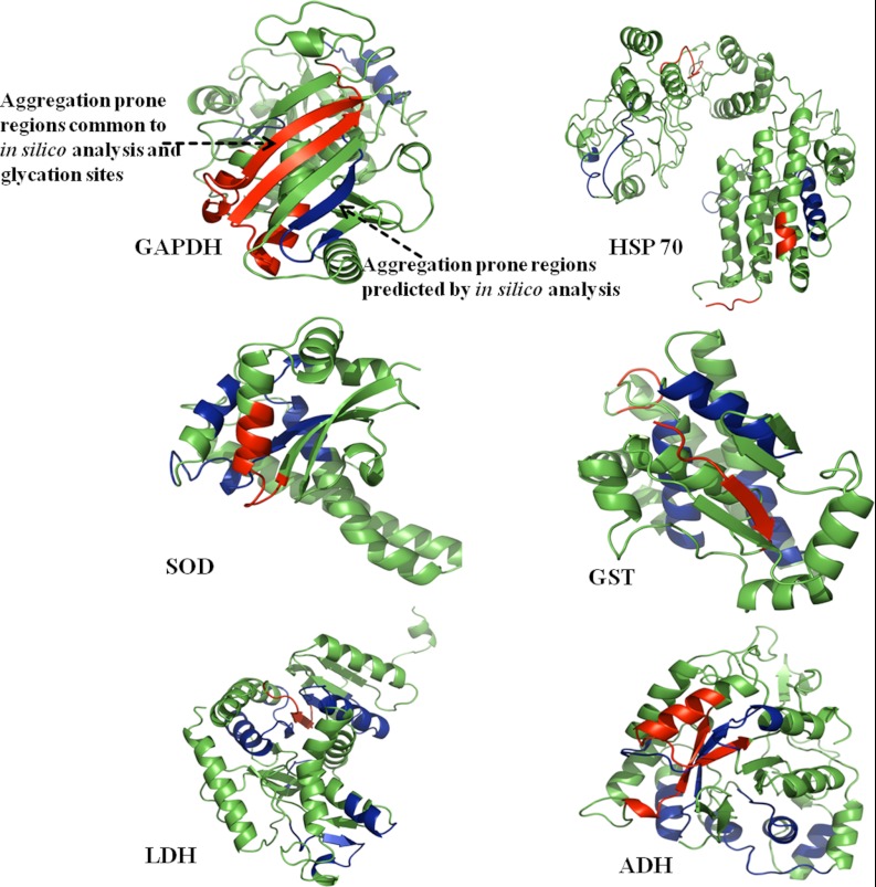
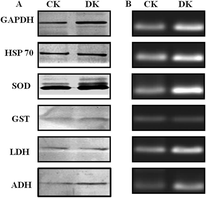
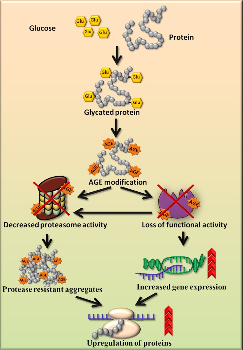
Glycation induced protein aggregation has been implicated in the development of diabetic complications and neurodegenerative diseases. These aggregates are known to be resistant to proteolytic digestion. Here we report the identification of protease resistant proteins from the streptozotocin induced diabetic rat kidney, which included enzymes in glucose metabolism and stress response proteins. These protease resistant proteins were characterized to be advanced glycation end products modified and ubiquitinated by immunological and mass spectrometry analysis. Further, diabetic rat kidney exhibited significantly impaired proteasomal activity. The functional analysis of identified physiologically important enzymes showed that their activity was reduced in diabetic condition. Loss of functional activity of these proteins was compensated by enhanced gene expression. Aggregation prone regions were predicted by in silico analysis and compared with advanced glycation end products modification sites. These findings suggested that the accumulation of protein aggregates is an inevitable consequence of impaired proteasomal activity and protease resistance due to advanced glycation end products modification.


Figures

Fig. 1.
Fig. 1.
In vitro evidence on protease resistance of glycated protein. BSA was glycated and ribosylated with glucose and ribose respectively, which showed protease resistance to tryptic digestion. Lane 2, 3, and 4 contains undigested BSA, Gly-BSA, and Rib-BSA respectively, whereas lane 5, 6, and 7 contains the same digested with trypsin, Try-BSA, Try-Gly-BSA, and Try-Rib-BSA respectively, and Lane 1 contains SDS-PAGE marker. The experiment was repeated independently three times.
Fig. 2.
Fig. 2.
AGE and ubiquitin modification of PRPs. The protein lysate of control (CK) and diabetic (DK) rat kidney was separated on SDS-PAGE with and without tryptic digestion. Western blot analysis was performed with anti-AGE or anti-ubiquitin antibodies for detection of PRPs with AGE and ubiquitin modification. A, SDS-PAGE: Lane 2 and 3 showing undigested control (CK) and diabetic (DK) rat kidney lysate whereas lane 4 and 5 contains the same digested with trypsin (Try-CK, Try-DK). The same samples were immunoblotted with Anti-AGE (B) and Anti-ubiquitin (C) antibody, respectively. The experiment was repeated independently three times.
Fig. 3.
Fig. 3.
Total protease and proteasomal activity. Control (CK) and diabetic (DK) rat kidney protein lysate was assessed for total protease activity by A, Azocasein assay and B, In- gel protease assay. These assays demonstrated that total protease activity is significantly reduced in DK. Formation of fluorescent AMC from AMC-tagged peptide was determined to assess (C) Proteasomal activity of rat kidney homogenate of CK and DK, and D, In vitro MGO modified proteasome activity. Bars represent means ± S.E. from three independent experiments. *p < 0.05.
Fig. 4.
Fig. 4.
Validation of LC-MSE identified PRPs by Western blotting. Protein lysate of Control (CK) and diabetic (DK) rat kidney was digested with trypsin, and separated on SDS-PAGE, followed by immunoblotting with antibodies against GAPDH, HSP 60, SOD, GST, LDH, and ADH. Presence of these proteins in DK after trypsin digestion confirmed the identification of PRPs by mass spectrometry. The experiment was repeated independently three times.
Fig. 5.
Fig. 5.
Functional assays of identified PRPs. The enzymatic activities of A, SOD, B, LDH, C, ADH, and D, GST were analyzed for control (CK) and diabetic (DK) rat kidney protein lysate using protocols described in supplemental method S1. In DK, significant reduction in enzymatic activity was observed. Bars represent means ± S.E. from three independent experiments. *p < 0.05.
Fig. 6.
Fig. 6.
Prediction of aggregation prone regions. Protein structures of rat GAPDH, HSP 60, SOD, GST, LDH and ADH were modeled by CPH 3.0 model server and analyzed using PyMol. Aggregation prone regions in these proteins were predicted in silico using Aggrescan, Tango, PASTA, and Waltz web servers (blue color). The regions common to aggregation prone sequences and glycation sites were highlighted in the red color, indicating that most of the glycation sites were overlapping with predicted aggregation hotspots.
Fig. 7.
Fig. 7.
Expression analysis of PRPs. A, Protein expression of GAPDH, HSP 60, SOD, GST, LDH, and ADH in control (CK) and diabetic (DK) rat kidney was measured by the Western blot analysis. B, The same proteins were analyzed at the transcript level by performing semi-quantitative RT-PCR using total RNA isolated from control and diabetic rat kidney tissues. All samples were analyzed on 2% agarose gels containing gel red. Both analyses showed up-regulation of PRPs in diabetic condition. Results shown are representative of three independent experiments.
Fig. 8.
Fig. 8.
Schematic diagram for accumulation of PRPs and up-regulation of proteins. The proposed model shows that AGE modification and impairment in proteasomal activity results in accumulation of protease resistant aggregates. AGE modification causes loss of functional activity of enzymes, which is compensated by increased gene expression. Both the inability of proteasome to remove protease resistant aggregates and decreased enzyme activity may cause up-regulation of proteins in the diabetic condition.

References

    1. Brownlee M. (2001) Biochemistry and molecular cell biology of diabetic complications. Nature. 414, 813–820 - PubMed
    1. Peppa M., Uribarri J., Vlassara H. (2003) Glucose, Advanced Glycation End products, and Diabetes complications: What is new and what works. Clin. Diabetes. 21, 186–187
    1. Bulteau A. L., Verbeke P., Petropoulos I., Chaffotte A. F., Friguet B. (2001) Proteasome inhibition in glyoxal-treated fibroblasts and resistance of glycated glucose-6-phosphate dehydrogenase to 20s proteasome degradation in vitro. J. Biol. Chem. 276, 45662–45668 - PubMed
    1. Monnier V. M., Kohn R. R., Cerami A. (1984) Accelerated age-related browning of human collagen in diabetes mellitus. Proc. Natl. Acad. Sci. U.S.A. 81, 583–587 - PMC - PubMed
    1. Reddy G. K., Stehno-Bittel L., Enwemeka C. S. (2002) Glycation-induced matrix stability in the rabbit achilles tendon. Arch. Biochem. Biophys. 399, 174–180 - PubMed

Publication types

LinkOut - more resources