. 2013 Jan 7;41(1):648-56.
doi: 10.1093/nar/gks1015. Epub 2012 Oct 31.

Structural asymmetry in the Thermus thermophilus RuvC dimer suggests a basis for sequential strand cleavages during Holliday junction resolution

Affiliations

Structural asymmetry in the Thermus thermophilus RuvC dimer suggests a basis for sequential strand cleavages during Holliday junction resolution

Luan Chen et al. Nucleic Acids Res. .

Abstract

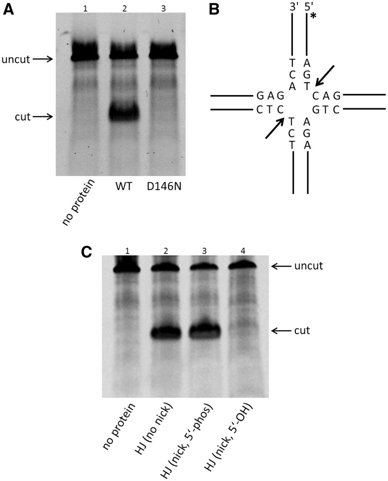
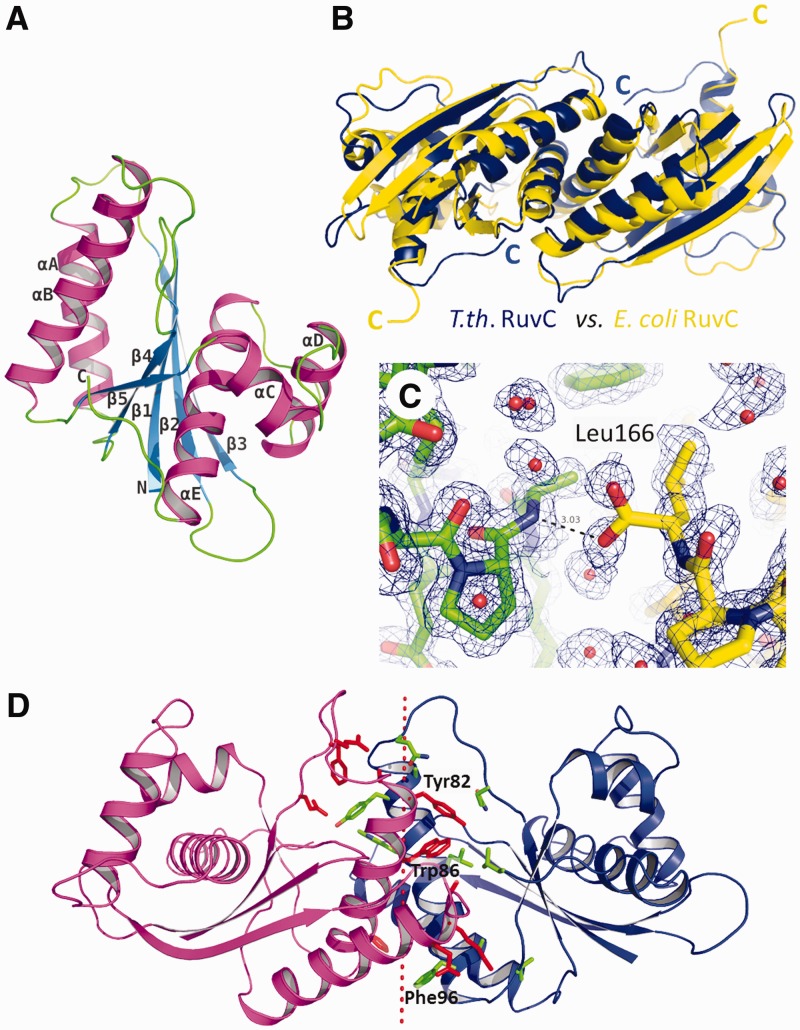
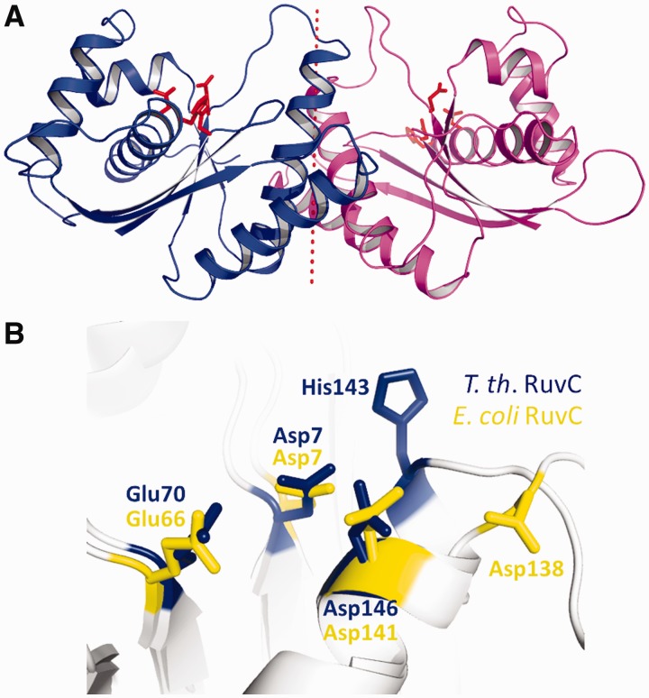
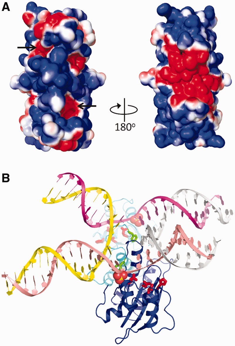
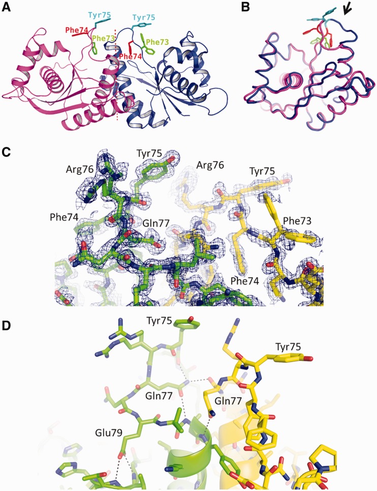
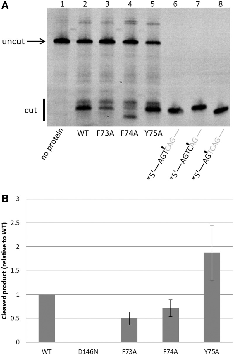
Holliday junction (HJ) resolvases are structure-specific endonucleases that cleave four-way DNA junctions (HJs) generated during DNA recombination and repair. Bacterial RuvC, a prototypical HJ resolvase, functions as homodimer and nicks DNA strands precisely across the junction point. To gain insights into the mechanisms underlying symmetrical strand cleavages by RuvC, we performed crystallographic and biochemical analyses of RuvC from Thermus thermophilus (T.th. RuvC). The crystal structure of T.th. RuvC shows an overall protein fold similar to that of Escherichia coli RuvC, but T.th. RuvC has a more tightly associated dimer interface possibly reflecting its thermostability. The binding mode of a HJ-DNA substrate can be inferred from the shape/charge complementarity between the T.th. RuvC dimer and HJ-DNA, as well as positions of sulfate ions bound on the protein surface. Unexpectedly, the structure of T.th. RuvC homodimer refined at 1.28 Å resolution shows distinct asymmetry near the dimer interface, in the region harboring catalytically important aromatic residues. The observation suggests that the T.th. RuvC homodimer interconverts between two asymmetric conformations, with alternating subunits switched on for DNA strand cleavage. This model provides a structural basis for the 'nick-counter-nick' mechanism in HJ resolution, a mode of HJ processing shared by prokaryotic and eukaryotic HJ resolvases.


Figures

Figure 1.
Figure 1.
T.th. RuvC functions as a HJ resolvase in vitro. (A) Denaturing polyacrylamide gel analysis of HJ-DNA resolution by the wild-type and the D146N mutant T.th. RuvC proteins. The top band corresponds to the fluorescently labeled uncut 64-base oligonucleotide (uncut), whereas the bottom band corresponds to the cleaved product (cut). The mutation of a catalytic residue Asp146 abolishes the DNA cleavage activity. (B) Structure of the ‘bi-mobile’ HJ substrate and the sequences near the junction point. The dominant cutting sites by the wild-type T.th. RuvC are indicated by arrows. The asterisk (*) indicates the position of the 6-FAM label. (C) Cleavage of unnicked or pre-nicked HJ substrates (5′-end at the nick having either phosphate or hydroxyl group) by the wild-type T.th. RuvC, analyzed on a denaturing gel. The result shows that resolution of the pre-nicked HJ requires presence of the 5′-phosphate group at the nick. All reactions shown in Figure 1 were carried out at a protein concentration of 500 nM and 100 nM of HJ-DNA substrate.
Figure 2.
Figure 2.
Structure of T.th. RuvC. (A) T.th. RuvC monomer shown in a ribbon diagram, with the secondary structure elements labeled. (B) T.th. RuvC dimer in blue superimposed on the E. coli RuvC dimer in yellow. The α-helices are packed more tightly at the dimer interface in the T.th. RuvC dimer, with the C-termini of the proteins tucked in. (C) The C-terminal residue Leu166 of T.th. RuvC is well ordered, with the carboxyl group involved in hydrogen bonding across the dimer interface. The two different molecules within the T.th. RuvC dimer are colored differently (yellow and green) to highlight the intermolecular interaction. The simulated annealing composite omit 2Fo-Fc map is contoured at 1.0σ. (D) Residues involved in the dimer interface are shown, with the aromatic residues Tyr82, Trp86 and Phe96 highlighted. The dotted line represents the pseudo 2-fold axis relating the two molecules in the T.th. RuvC dimer.
Figure 3.
Figure 3.
The enzyme active site of T.th. RuvC. (A) Ribbon diagram of the T.th. RuvC dimer, with the catalytic residues Asp7, Glu70, His143 and Asp146 shown in red sticks to indicate the location of the active sites. (B) Superposition of the catalytic residues from E. coli RuvC (yellow) and T.th. RuvC (blue).
Figure 4.
Figure 4.
Surface potential of the T.th. RuvC dimer and a possible mode of HJ-DNA binding. (A) Solvent accessible surface of the T.th. RuvC dimer colored according to the electrostatic surface potential (red: −1 kT/e to blue: +1 kT/e), shown in two orientations rotated by 180°. The location of the active sites is indicated by arrows. (B) A hypothetical model of T.th. RuvC dimer bound to a HJ-DNA. The spheres represent sulfate ions bound on the protein surface (in the crystal form II), whereas the active site residues are shown in red sticks. The Phe73 side chains are shown by green sticks. A close-up view around the junction point is shown in Supplementary Figure S7.
Figure 5.
Figure 5.
The asymmetric loop. (A) Ribbon diagram of the T.th. RuvC dimer, with the aromatic residues in the asymmetric loop region Phe73, Phe74 and Tyr75 shown in sticks. (B) Superposition of the two T.th. RuvC molecules from the asymmetric dimer, with Phe73, Phe74 and Tyr75 colored as in (A). The loop region with the greatest conformational difference between the two molecules is highlighted by an arrow. Deviations of the Cα positions in this superposition are shown in Supplementary Figure S6. (C) Electron density for the asymmetric loops. Simulated annealing composite omit 2Fo-Fc map within 1.7 Å from the protein atoms is shown, contoured at 1.0σ. Arg76 from one of the molecules and Phe73 from the other molecule show multiple conformations. (D) Hydrogen-bonding network involving Gln77 and Glu79 stabilizes the asymmetric conformations of the loops. The hydrogen bonds are indicated by the dotted lines.
Figure 6.
Figure 6.
HJ-DNA resolution by the T.th. RuvC mutants. (A) Resolution of the unnicked HJ-DNA substrate by the wild-type and mutant T.th. RuvC proteins, analyzed on a denaturing gel. Mutations of the aromatic residues in the asymmetric loop region differently affect the HJ resolvase activity. Lanes 6–8 have fluorescently labeled oligonucleotides corresponding to cleaved products, as size markers. The F74A mutant (lane 4) shows a distinct pattern with unique sequence selectivity in DNA cleavage. The enzyme and HJ-DNA substrate concentration was 500 and 100 nM, respectively. (B) Quantification of the total products generated, normalized against the wild-type. The error bars show standard deviations between three experiments.

References

    1. West SC. Molecular views of recombination proteins and their control. Nat. Rev. Mol. Cell Biol. 2003;4:435–445. - PubMed
    1. Holliday R. A mechanism for gene conversion in fungi. Genet. Res. 1964;5:282–304. - PubMed
    1. Lilley DM, White MF. The junction-resolving enzymes. Nat. Rev. Mol. Cell Biol. 2001;2:433–443. - PubMed
    1. Michel B, Boubakri H, Baharoglu Z, LeMasson M, Lestini R. Recombination proteins and rescue of arrested replication forks. DNA Repair. 2007;6:967–980. - PubMed
    1. Biertümpfel C, Yang W, Suck D. Crystal structure of T4 endonuclease VII resolving a Holliday junction. Nature. 2007;449:616–620. - PubMed

Publication types