. 2012 Nov-Dec;13(6):720-7.
doi: 10.3348/kjr.2012.13.6.720. Epub 2012 Oct 12.

Radiation dose reduction of chest CT with iterative reconstruction in image space - Part II: assessment of radiologists' preferences using dual source CT

Affiliations

Radiation dose reduction of chest CT with iterative reconstruction in image space - Part II: assessment of radiologists' preferences using dual source CT

Hye Jeon Hwang et al. Korean J Radiol. 2012 Nov-Dec.

Abstract

Objective: To evaluate the impact of radiation dose and reconstruction algorithms on radiologists' preferences, and whether an iterative reconstruction in image space (IRIS) can be used for dose reduction in chest CT.

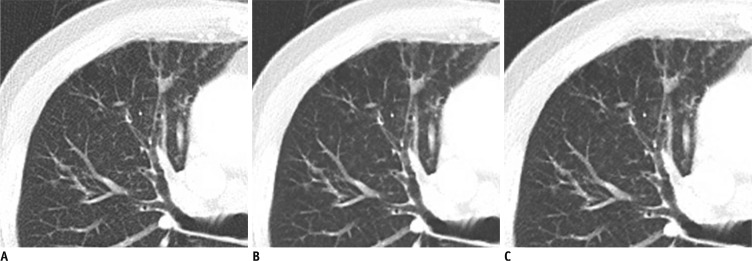
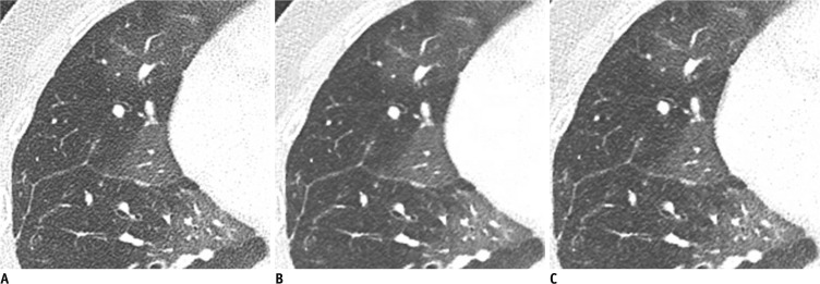
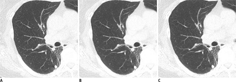
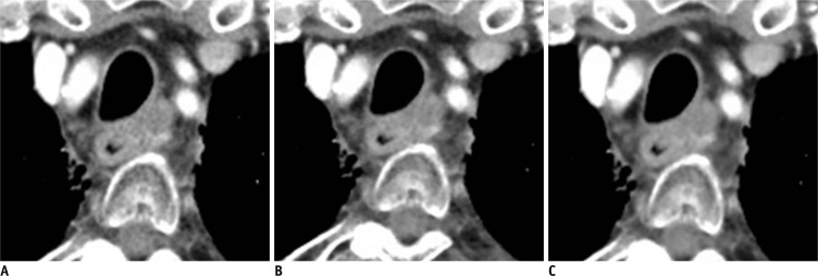
Materials and methods: Standard dose chest CT (SDCT) in 50 patients and low dose chest CT (LDCT) in another 50 patients were performed, using a dual-source CT, with 120 kVp and same reference mAs (50 mAs for SDCT and 25 mAs for LDCT) employed to both tubes by modifying the dual-energy scan mode. Full-dose data were obtained by combining the data from both tubes and half-dose data were separated from one tube. These were reconstructed by using a filtered back projection (FBP) and IRIS: full-dose FBP (F-FBP); full-dose IRIS (F-IRIS); half-dose FBP (H-FBP) and half-dose IRIS (H-IRIS). Ten H-IRIS/F-IRIS, 10 H-FBP/H-IRIS, 40 F-FBP/F-IRIS and 40 F-FBP/H-IRIS pairs of each SDCT and LDCT were randomized. The preference for clinical usage was determined by two radiologists with a 5-point-scale system for the followings: noise, contrast, and sharpness of mediastinum and lung.

Results: Radiologists preferred IRIS over FBP images in the same radiation dose for the evaluation of the lungs in both SDCT (p = 0.035) and LDCT (p < 0.001). When comparing between H-IRIS and F-IRIS, decreased radiation resulted in decreased preference. Observers preferred H-IRIS over F-FBP for the lungs in both SDCT and LDCT, even with reduced radiation dose by half in IRIS image (p < 0.05).

Conclusion: Radiologists' preference may be influenced by both radiation dose and reconstruction algorithm. According to our preliminary results, dose reduction at 50% with IRIS may be feasible for lung parenchymal evaluation.

Keywords: Chest CT; Iterative reconstruction in image space; Preference; Radiation dose reduction.


Figures

Fig. 1
Fig. 1
Standard dose contrast enhanced-chest CT in 77-year-old man (BMI 22 kg/m2) with sequalae of previous inflammation in right upper lobe. A-C. Transverse CT images at 5 mm thickness with full radiation dose reconstructed with (A) filtered back projection (FBP) and (B) image reconstruction in image space (IRIS), and (C) with half radiation dose reconstructed with IRIS. When two readers compared F-FBP (A) and F-IRIS (B) images as reconstruction effect, their preference scores were 5 and 4 for lung 5 mm images, 2 and 3 for mediastinum, 5 and 3 for lung 1 mm images, and 5 and 4 for overall images (not shown). When two readers compared F-FBP (A) and H-IRIS (C) images as reconstruction effect and radiation dose effect, readers' preference scores were 4 and 3 for lung 5 mm images, 3 and 3 for mediastinum, 4 and 4 for lung 1 mm images, and 4 and 3 for overall images (not shown). BMI = body mass index, F-FBP = full dose image with filtered back projection, F-IRIS = full dose image with iterative reconstruction in image space, H-IRIS = half dose image with iterative reconstruction in image space
Fig. 2
Fig. 2
Standard dose contrast enhanced-chest CT in 19-year-old man (BMI, 21 kg/m2) with pulmonary hypertension. A-C. Transverse CT images at 1 mm thickness with full radiation dose reconstructed with (A) FBP and (B) IRIS, and (C) with half radiation dose reconstructed with IRIS. When two readers compared F-FBP (A) and F-IRIS (B) images, reconstruction effect, they both had preference scores of 4 and 4 for lung 5 mm images, 3 and 3 for mediastinum, 4 and 3 for lung 1 mm images and 3 and 4 for overall (not shown). When two readers compared F-FBP (A) and H-IRIS (C) images, as reconstruction effect and radiation dose effect, readers' preference scores were 4 and each for lung 5 mm images, 3 and 2 each for mediastinum, 4 and 3 for lung 1 mm images and 4 and 3 for overall (not shown). BMI = body mass index, F-FBP = full dose image with filtered back projection, F-IRIS = full dose image with iterative reconstruction in image space, H-IRIS = half dose image with iterative reconstruction in image space
Fig. 3
Fig. 3
Low dose chest CT in 62-year-old woman (BMI, 27 kg/m2) with postoperative lung cancer. Transverse CT images at 5 mm thickness with full radiation dose reconstructed with (A) FBP and (B) IRIS, and (C) with half radiation dose reconstructed with IRIS. When two readers compared F-FBP (A) and F-IRIS (B) images as reconstruction effect, readers' preference scores of 4 and 4 for lung 5 mm images, 3 and 2 for mediastinum, 5 and 4 for lung 1 mm images, and 4 and 4 for overall (not shown). When they compared F-FBP (A) and H-IRIS (C) as reconstruction effect and radiation dose effect, two readers had preference scores of 4 and 4 each for lung 5 mm images, 2 and 3 for mediastinum, 5 and 4 for lung 1 mm images, and 4 and 4 overall (not shown). BMI = body mass index, F-FBP = full dose image with filtered back projection, F-IRIS = full dose image with iterative reconstruction in image space, H-IRIS = half dose image with iterative reconstruction in image space
Fig. 4
Fig. 4
Standard dose contrast enhanced-chest CT in 77-year-old man (BMI, 22 kg/m2) with esophageal cancer. Transverse CT image at 5 mm thickness with full radiation dose reconstructed with (A) FBP and (B) IRIS, and (C) with half radiation dose reconstructed with IRIS. When two readers compared F-FBP (A) and F-IRIS (B) images, as reconstruction effect, readers' preference scores were 2 and 2 each for mediastinum, 4 and 3 for lung 5 mm images, 4 and 3 for lung 1 mm images and 4 and 3 for overall (not shown). When they compared F-FBP (A) and H-IRIS (C) as reconstruction effect and radiation dose effect, readers had preference scores of 2 and 2 each for mediastinum, 4 and 4 for lung 5 mm/1 mm images, and 3 overall (not shown). BMI = body mass index, F-FBP = full dose image with filtered back projection, F-IRIS = full dose image with iterative reconstruction in image space, H-IRIS = half dose image with iterative reconstruction in image space

References

    1. Rogers LF. Dose reduction in CT: how low can we go? AJR Am J Roentgenol. 2002;179:299. - PubMed
    1. Yu L, Li H, Fletcher JG, McCollough CH. Automatic selection of tube potential for radiation dose reduction in CT: a general strategy. Med Phys. 2010;37:234–243. - PubMed
    1. Greess H, Wolf H, Baum U, Lell M, Pirkl M, Kalender W, et al. Dose reduction in computed tomography by attenuation-based on-line modulation of tube current: evaluation of six anatomical regions. Eur Radiol. 2000;10:391–394. - PubMed
    1. Schenzle JC, Sommer WH, Neumaier K, Michalski G, Lechel U, Nikolaou K, et al. Dual energy CT of the chest: how about the dose? Invest Radiol. 2010;45:347–353. - PubMed
    1. Lell MM, May M, Deak P, Alibek S, Kuefner M, Kuettner A, et al. High-pitch spiral computed tomography: effect on image quality and radiation dose in pediatric chest computed tomography. Invest Radiol. 2011;46:116–123. - PubMed

Publication types

LinkOut - more resources