

Opportunities for 2-[(18)F] fluoro-2-deoxy-D-glucose PET/CT in cervical-vaginal neuroendocrine carcinoma: case series and literature review
- PMID: 23118575
- PMCID: PMC3484297
- DOI: 10.3348/kjr.2012.13.6.760
Opportunities for 2-[(18)F] fluoro-2-deoxy-D-glucose PET/CT in cervical-vaginal neuroendocrine carcinoma: case series and literature review
Abstract
Objective: Neuroendocrine cervical carcinoma is a rare subtype of cervical cancer. These tumors exhibit an aggressive behavior with early regional lymph node and distant metastases. The purpose of our study was to describe five cases of neuroendocrine cervical-vaginal carcinoma and to discuss the potential of the 2-[(18)F] fluoro-2-deoxy-D-glucose positron emission tomography/computed tomography ((18)F-FDG PET/CT) scan for the detection of this rare malignancy.
Materials and methods: Five cases of cervical-vaginal neuroendocrine tumor were retrospectively collected, during a two year (from September 2009 to August 2011) period in our hospital. The clinical staging distributions were International Federation of Gynecology and Obstetrics (FIGO) stage IB2 (1 of 5), stage IIA (3 of 5) and stage IVA (1 of 5).
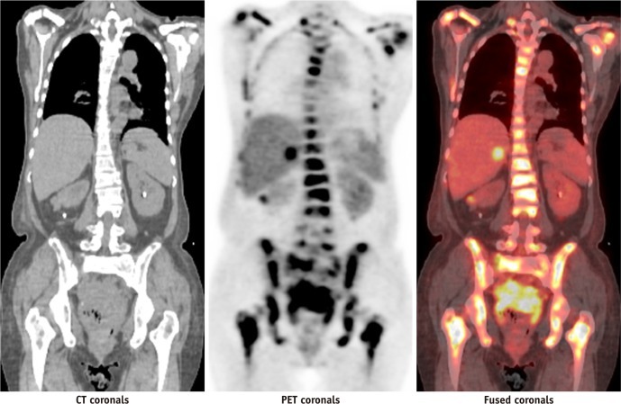
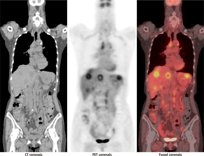
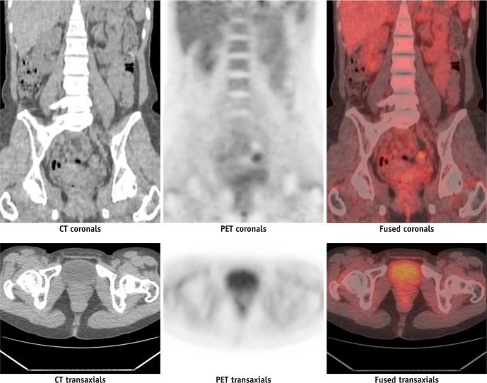
Results: Two cases (cases 1 and 4) were restaged after (18)F-FDG PET/CT scan in the initial staging process. Post-treatment (18)F-FDG PET/CT scans, in three patients, revealed positive findings for tumor recurrence or lymph node metastases. Two patients (cases 2 and 3) died of tumor within two years.
Conclusion: (18)F-FDG PET/CT scan is a useful tool in cervical-vaginal neuroendocrine tumor. In its initial staging, the (18)F-FDG PET/CT scan may help assess the possible nodal involvement or early hematogeneous spreading. We can also use the (18)F-FDG PET/CT to detect local recurrence and to evaluate the treatment response after clinical manipulation.
Keywords: 18F-FDG PET/CT; Cervical carcinoma; Neuroendocrine carcinoma.
Figures


References
-
- Oberg K, Castellano D. Current knowledge on diagnosis and staging of neuroendocrine tumors. Cancer Metastasis Rev. 2011;30(Suppl 1):3–7. - PubMed
-
- Albores-Saavedra J, Gersell D, Gilks CB, Henson DE, Lindberg G, Santiago H, et al. Terminology of endocrine tumors of the uterine cervix: results of a workshop sponsored by the College of American Pathologists and the National Cancer Institute. Arch Pathol Lab Med. 1997;121:34–39. - PubMed
-
- Sato Y, Shimamoto T, Amada S, Asada Y, Hayashi T. Large cell neuroendocrine carcinoma of the uterine cervix: a clinicopathological study of six cases. Int J Gynecol Pathol. 2003;22:226–230. - PubMed
-
- Krivak TC, McBroom JW, Sundborg MJ, Crothers B, Parker MF. Large cell neuroendocrine cervical carcinoma: a report of two cases and review of the literature. Gynecol Oncol. 2001;82:187–191. - PubMed
-
- Reig Castillejo A, Membrive Conejo I, Foro Arnalot P, Rodríguez de Dios N, Algara López M. Neuroendocrine small cell carcinoma of the uterine cervix. Clin Transl Oncol. 2010;12:512–513. - PubMed
