. 2012:2012:156563.
doi: 10.1155/2012/156563. Epub 2012 Oct 14.

Effects of Vitamin D Supplementation during the Induction and Progression of Osteoarthritis in a Rat Model

Affiliations

Effects of Vitamin D Supplementation during the Induction and Progression of Osteoarthritis in a Rat Model

E C Castillo et al. Evid Based Complement Alternat Med. 2012.

Abstract

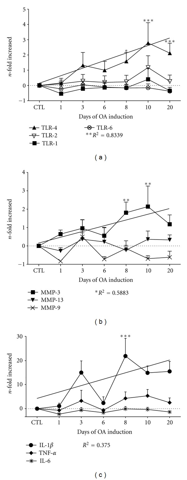
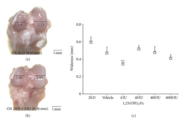
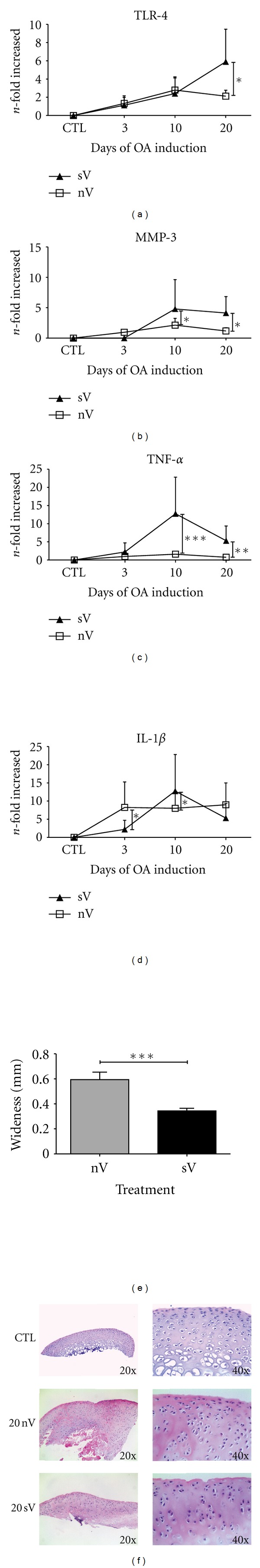
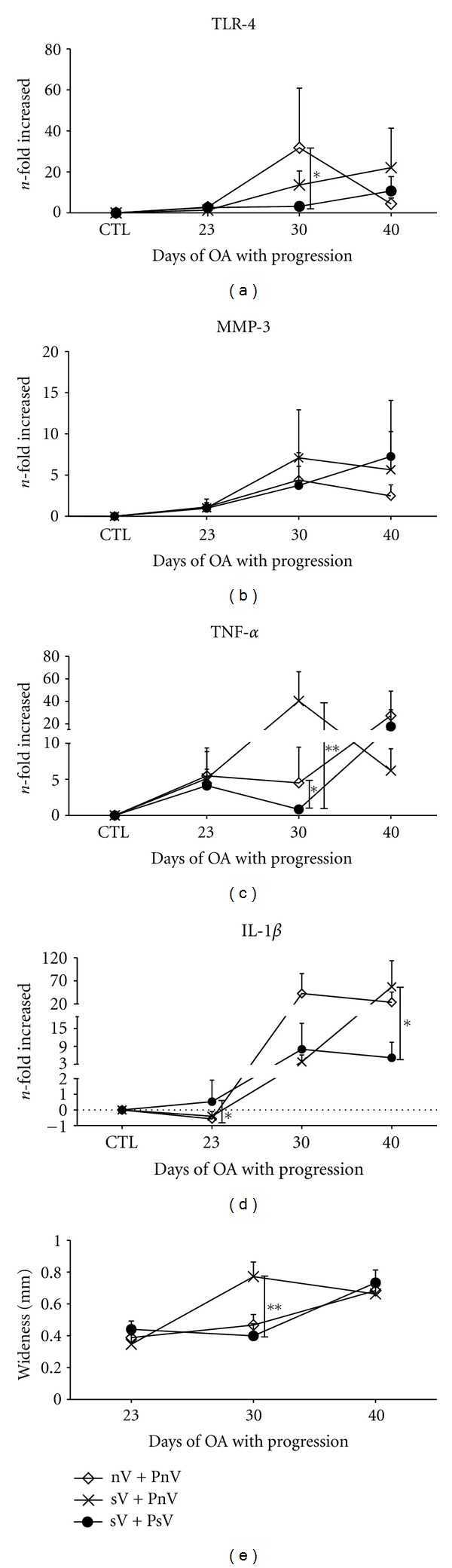
Epidemiological studies correlate low levels of vitamin D with the osteoarthritis (OA) progression. Cytokines and metalloproteases play a major role in OA promoting the inflammation and degradation of the cartilage and can be induced through the Toll-like receptor (TLR) pathway. The aim of this study was to evaluate the protective effect of vitamin D supplementation on the development of osteoarthritis (OA) through examining the genetic regulation of TLRs, cytokines, and metalloproteases in chondrocytes as well as the wideness of cartilage in rats with OA. Our results demonstrate that the signaling through TLR-4 is a proinflammatory mechanism in osteoarthritis that drives the upregulation of MMP-3, IL-1β, and TNF-α gene expression, leading to cartilage degradation and inflammation. Vitamin D supplementation had a protective effect during the onset but not during the chronic stage of OA in the rat model.


Figures

Figure 1
Figure 1
Experimental groups. A schematic example of the 20-day induction group and the 40-day progression group of rats with OA with or without supplementation is shown. The same scheme was applied for the other induction (3 and 10 days) and progression (20 and 40 days) groups. Vitamin D (4 IU/kg/d) was administrated orally with an esophagogastric cannula 14 HALO. The arrow indicates the group without vitamin supplementation (nV); the pointed arrow indicates the supplementation with vitamin D (sV). Each group was conformed by 10 rats, and every experiment was performed at least by triplicate.
Figure 2
Figure 2
TLR-4, MMP-3, IL-1β, and TNF-α expression was induced during osteoarthritis induction. Chondrocytes were collected and assayed for gene expression by real-time RT-PCR. The relative expression levels were normalized to the expression of the housekeeping GAPDH gene expression and presented as the n-fold difference. (a) TLR-2 and TLR-1 showed a similar pattern of expression during OA induction, whereas TLR-6 was only expressed during the acute stage of OA. TLR-4 showed a remarkable increase in expression with respect to the CTL group throughout OA induction, with a significant linear trend (R 2 = 0.8339; P = 0.0041). (b) The relative expression levels of MMP-3, MMP-9, and MMP-13, normalized to GAPDH gene expression, are shown. MMP-3 expression increased during OA induction (R 2 = 0.5883, P = 0.0219) and was highest on days 8 (1.8-fold; 95% CI 0.005241 to 3.609) and 10 (2.1-fold; 95% CI 0.3375 to 3.941). (c) The relative expression of IL-1β, IL-6, and TNF-α, normalized to GAPDH gene expression, is shown. IL-1β gene expression increased during OA induction (R 2 = 0.375), while TNF-α gene expression changed modestly (R 2 = 0.4173), with the highest increase on day 10 (1.6-fold). The results are pooled data from five independent experiments with 50 total rats. The asterisks indicate *P < 0.05, **P < 0.01, and ***P < 0.001, which were determined using a 2-way ANOVA followed by Tukey's post hoc test to compare the experimental groups with the CTL group. The error bars indicate the SEM.
Figure 3
Figure 3
Vitamin D reduces the wideness of OA condyles. (a) A representative image of the femoral condyles from a rat with 20 days of OA without treatment. (b) A representative image of the femoral condyles from a rat treated with 4 IU/kg/day of vitamin D. The difference in wideness was taken as an indicator of the degree of severity of OA hypertrophy. (c) The graph presents the average wideness at a given dose from 3 different experiments with 15 total rats. A decrease in the severity of 0.34 ± 0.047 mm (95% CI 0.05367 to 0.4459, **P < 0.01) was determined using a one-way ANOVA with Dunnet's post hoc test to compare all of the conditions with the control group (OA of 20 days without vitamin D). The error bars indicate the SEM. The scale bar = 1 mm 8x.
Figure 4
Figure 4
The protective effect of vitamin D is independent of the transcriptional modulation of TLR-4. The evaluation was performed during the OA induction period for (a)–(d). The gene expression of TLR4 (a), MMP-3 (b), TNF-α (c), and IL-1β (d) from rats in the nV group (□) compared with the sV group (▲) is shown. The gene expression levels were normalized to GAPDH gene expression and presented as the n-fold difference in expression. (e) A comparison of the hypertrophy (wideness) of the condyles from rats with 20 days of OA that were supplemented with vitamin D (sV) to that of rats with no supplementation (nV). (f) A morphological comparison between condyles of control rats and rats with 20 days of OA induction treated with vitamin D (sV) and nontreated (nV) by HE stain. The supplementation was performed daily with 4 IU of vitamin D. The results are expressed as pooled data from three independent experiments with 16 to 18 total rats (a–d) or with 6 rats per group (e). The error bars indicate the SEM. A t-test with an F test was performed to obtain the indicated statistical significance values: P < 0.05, **P < 0.01, and ***P < 0.001. Abbreviations indicate the following treatments: nV (without vitamin supplementation); sV (with vitamin supplementation).
Figure 5
Figure 5
Vitamin D has no effect on OA severity. The evaluation of OA was performed during the OA progression phase.(a)–(d). The gene expression of TLR-4 (a), MMP-3 (b), TNF-α (c), and IL-1β (d) from rats in the sV + Psv group (●) compared with the sV + PnV (×) and the nV + PnV () groups. The gene expression levels were normalized to GAPDH gene expression and are presented as the n-fold difference in expression. (e) A comparison of the hypertrophy (wideness) of condyles from the rats of the sV + PsV (●), sV + PnV (×) and nV + PnV () groups is shown. The results shown are pooled data from three independent experiments with 16 to 18 total rats (a–d) or with 12 to 16 total rats (e). The error bars indicate the SEM. A t-test with an F test was performed; the notations indicate *P < 0.05 and **P < 0.01. The following groups were included: sV + PsV (vitamin supplementation during induction and progression with supplementation of vitamin); sV + PnV (vitamin supplementation during induction and progression without vitamin supplementation); nV + PnV (without vitamin supplementation during induction and progression without vitamin supplementation). For the supplement, 4 IU of vitamin D was administrated daily.

References

    1. Sadeghi K, Wessner B, Laggner U, et al. Vitamin D3 down-regulates monocyte TLR expression and triggers hyporesponsiveness to pathogen-associated molecular patterns. European Journal of Immunology. 2006;36(2):361–370. - PubMed
    1. Haroon M, Bond U, Quillinan N, Phelan MJ, Regan MJ. The prevalence of vitamin D deficiency in consecutive new patients seen over a 6-month period in general rheumatology clinics. Clinical Rheumatology. 2011;30(6):789–794. - PubMed
    1. Bischoff-Ferrari HA, Zhang Y, Kiel DP, Felson DT. Positive association between serum 25-hydroxyvitamin D level and bone density in osteoarthritis. Arthritis & Rheumatism. 2005;53(6):821–826. - PubMed
    1. Heidari B, Heidari P, Hajian-Tilaki K. Association between serum vitamin D deficiency and knee osteoarthritis. International Orthopaedics. 2011;35(11):1627–1631. - PMC - PubMed
    1. Chaganti RK, Parimi N, Cawthon P, Dam TL, Nevitt MC, Lane NE. Association of 25-hydroxyvitamin D with prevalent osteoarthritis of the hip in elderly men: the osteoporotic fractures in men study. Arthritis and Rheumatism. 2010;62(2):511–514. - PMC - PubMed

LinkOut - more resources