. 2012;7(10):e46962.
doi: 10.1371/journal.pone.0046962. Epub 2012 Oct 31.

GFP's mechanical intermediate states

Affiliations

GFP's mechanical intermediate states

John Saeger et al. PLoS One. 2012.

Abstract

Green fluorescent protein (GFP) mutants have become the most widely used fluorescence markers in the life sciences, and although they are becoming increasingly popular as mechanical force or strain probes, there is little direct information on how their fluorescence changes when mechanically stretched. Here we derive high-resolution structural models of the mechanical intermediate states of stretched GFP using steered molecular dynamics (SMD) simulations. These structures were used to produce mutants of EGFP and EYFP that mimic GFP's different mechanical intermediates. A spectroscopic analysis revealed that a population of EGFP molecules with a missing N-terminal α-helix was significantly dimmed, while the fluorescence lifetime characteristic of the anionic chromophore state remained unaffected. This suggests a mechanism how N-terminal deletions can switch the protonation state of the chromophore, and how the fluorescence of GFP molecules in response to mechanical disturbance might be turned off.


Conflict of interest statement

Competing Interests: The authors have declared that no competing interests exist.

Figures

Figure 1
Figure 1. The main mechanical intermediates of GFP.
The extension versus time graph for constant force pulls of GFP hydrated in a box of explicit water as derived by steered molecular dynamics simulations (SMD). Structures of the GFP molecule in significant mechanical intermediate states are shown. The major intermediates Corked, Barrel, and Ripped can be seen to have extended horizontal plateau regions compared to the others. The locations of the N-terminal deletions are indicated.
Figure 2
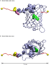
Figure 2. Transitioning into the Corked state.
A, B. Two views of the equilibrium structure. C, D. Two views of the Corked state at 50 pN. The hydrophobic “cork” consisting of Leu7 and Phe8 has rotated in the hydrophobic pocket somewhat but remains bound. The mechanism is suggestive of a peptide “lever” with a hydrophobic “fulcrum.” GFP variants in this state likely remain significantly fluorescent (see text).
Figure 3
Figure 3. Transitioning into the Barrel state.
A, B. Two views of the Barrel state at 100 pN. The “cork” consisting of Leu7 and Phe8 has been extracted from the hydrophobic pocket, and the hydrophobic pocket is significantly damaged. Nevertheless when pulled into this state, depending on the GFP variant, the molecule may retain significant fluorescence, or it may become almost completely dark (see text).
Figure 4
Figure 4. Transitioning into the Ripped states.
A. The initial structure in a space-filling representation prior to pulling, with the β1 (green) strand nestled between the darker colored β6 (blue) and β2 (red) strands. The N-terminal α-helix is colored to illustrate the N-terminal deletions (yellow (Δ5) and orange (Δ8)). B. A partially unpeeled β1 strand leaving behind a gap between strands β6 and β2 through which the chromophore (green) can be seen. C. A completely extracted β1 strand with the β6 and β2 strands resealed. D. The initial configuration in the ribbon representation with the β1 strand colored in green. The amino acid residues that form new contacts are shown in the space filling representation. E. The resealed barrel. F. The distances between the atoms involved in hydrogen bond formation from the pull at 200 pN that best demonstrated resealing behavior. Contacts between Gly31-Asn121 as well as Gly33-Leu119 are more stable than the contacts between Val29-Ile123. G. The one case where the barrel did not reseal. However, there is a remaining stable “mechanical core” shown in red that bears a structural resemblance to an intermediate state of the first type III module from human fibronectin . H. The first human fibronectin type III module (FnIII-1) in the intermediate state .
Figure 5
Figure 5. Absorption spectroscopy to probe for chromophore formation and relative brightness of the GFP mutants.
A. Absorption spectra of the non-mutated proteins. B. Absorption spectra of the mutants. C, D. Absorption spectra of the mutants (zoomed). In addition to the absorption peak in the visible band, a weaker second peak is seen in the vicinity of 390 nm for all mutants, which corresponds to the absorption band of a neutral chromophore. Note the UV absorption peak in the Δ22 mutants. This indicates that a complete 11-stranded β-barrel is not needed to form a chromophore. The shapes of the absorption spectra displayed by the EGFP and EYFP Δ5 mutants are similar to the absorption spectra of the wild type molecules. This indicates that the Δ5 mutants are largely anionic similar to the wild type molecules and are therefore bright. The EGFP Δ8 mutant displayed no significant absorption peak in the visible band and is therefore very dim. The EYFP Δ8 mutant in addition to an absorption peak in the UV displayed a significant absorption peak in the visible band and is therefore only slightly dim. The Δ22 mutants displayed no significant absorption peak in the visible band and are therefore dark. The baseline function was fit to the background as described in the methods.
Figure 6
Figure 6. Emission spectroscopy to probe for changes in the β-barrels of the GFP mutants.
A, B. Emission spectra of EGFP and EYFP based molecules excited at 460 nm and 480 nm respectively. Intensities were normalized to the absorbance of the molecules at 280 nm. The Δ22 mutants are seen to be completely dark when excited in the visible band. No significant changes in peak wavelength that would indicate changes to the structure of the β-barrel were observed . C, D. Fluorescence lifetime decay curves of EGFP and EYFP based molecules. Intensities were normalized at a time approximately 1 ns after the laser pulse. No significant changes in fluorescence lifetime that would indicate changes to the structure of the β-barrel were observed .
Figure 7
Figure 7. Correspondence of GFP's three major mechanical intermediate states between AFM and SMD .
The measurements of distance changes between mechanical intermediate states with SMD agreed with AFM measurements within the uncertainties of the measurements. The pattern of decreasing brightness of the mutant EGFP and EYFP molecules which were designed to mimic the structure of the molecules in the significant mechanical intermediate states were consistent with the reduced thermodynamic stability of these states measured by AFM . As we did not detect a significant energy barrier between the native state and the SMD corked state, we show the Corked state correlated with the bottom of the first energy well. The SMD Barrel state corresponds to the AFM GFPΔα state where the N-terminal α-helix has been pulled away from GFP's β-barrel. The SMD Ripped state corresponds to the AFM GFPΔαΔβ state where β1 strand has been pulled out of the β-barrel. The free energy diagram is adapted from Dietz and Rief . A summary of predicted fluorescence properties of the mechanical intermediate states based on measurements of the deletion mutants is also shown.

References

    1. Chalfie M, Tu Y, Euskirchen G, Ward WW, Prasher DC (1994) Green fluorescent protein as a marker for gene expression. Science 263: 802–805. - PubMed
    1. Lemmon CA, Ohashi T, Erickson HP (2011) Probing the Folded State of Fibronectin Type III Domains in Stretched Fibrils by Measuring Buried Cysteine Accessibility. J Biol Chem 286: 26375–26382. - PMC - PubMed
    1. Smith ML, Gourdon D, Little WC, Kubow KE, Eguiluz RA, et al. (2007) Force-induced unfolding of fibronectin in the extracellular matrix of living cells. PLoS Biol 5: e268. - PMC - PubMed
    1. Baneyx G, Baugh L, Vogel V (2002) Fibronectin extension and unfolding within cell matrix fibrils controlled by cytoskeletal tension. Proc Natl Acad Sci U S A 99: 5139–5143. - PMC - PubMed
    1. Brown AE, Discher DE (2009) Conformational changes and signaling in cell and matrix physics. Curr Biol 19: R781–789. - PMC - PubMed

Publication types

Substances

Associated data