. 2012;7(10):e48266.
doi: 10.1371/journal.pone.0048266. Epub 2012 Oct 30.

Cellular and molecular effect of MEHP Involving LXRα in human fetal testis and ovary

Affiliations

Cellular and molecular effect of MEHP Involving LXRα in human fetal testis and ovary

Vincent Muczynski et al. PLoS One. 2012.

Abstract

Background: Phthalates have been shown to have reprotoxic effects in rodents and human during fetal life. Previous studies indicate that some members of the nuclear receptor (NR) superfamilly potentially mediate phthalate effects. This study aimed to assess if expression of these nuclear receptors are modulated in the response to MEHP exposure on the human fetal gonads in vitro.

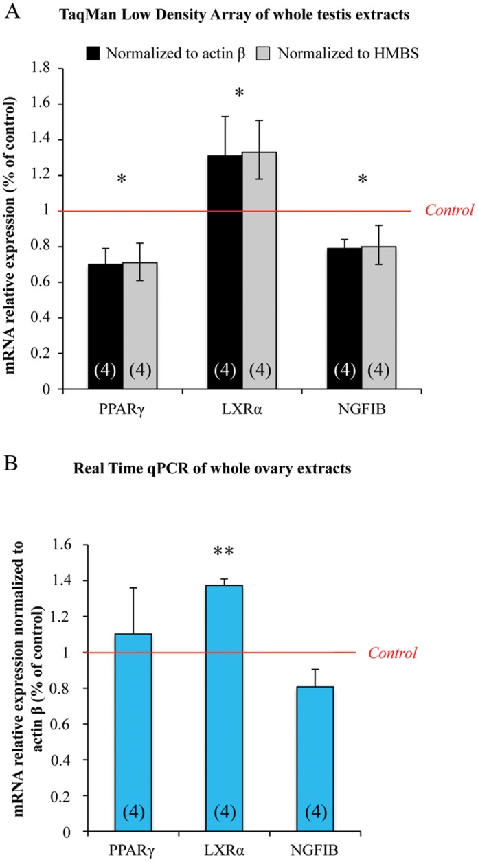
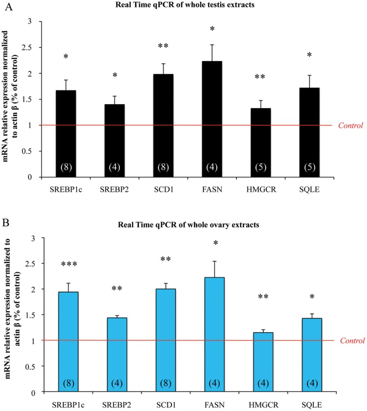
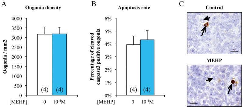
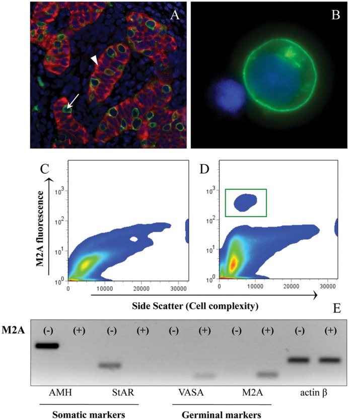
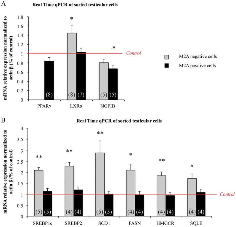
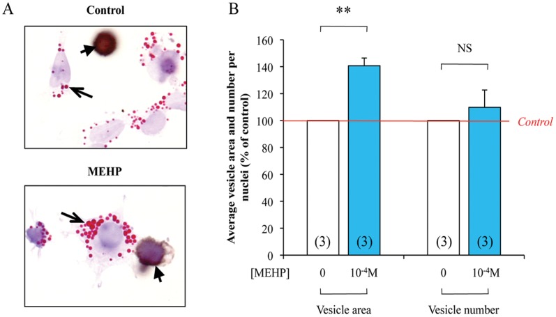
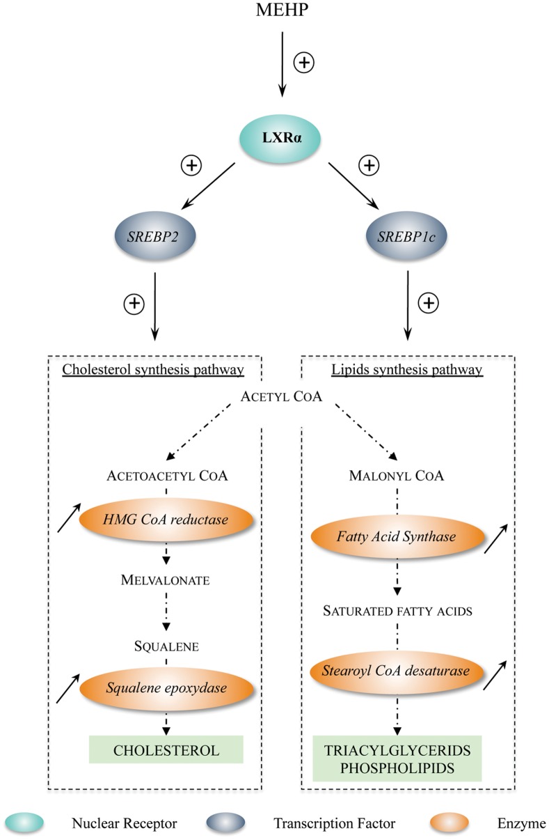
Methodology/principal findings: Testes and ovaries from 7 to 12 gestational weeks human fetuses were exposed to 10(-4)M MEHP for 72 h in vitro. Transcriptional level of NRs and of downstream genes was then investigated using TLDA (TaqMan Low Density Array) and qPCR approaches. To determine whether somatic or germ cells of the testis are involved in the response to MEHP exposure, we developed a highly efficient cytometric germ cell sorting approach. In vitro exposure of fetal testes and ovaries to MEHP up-regulated the expression of LXRα, SREBP members and of downstream genes involved in the lipid and cholesterol synthesis in the whole gonad. In sorted testicular cells, this effect is only observable in somatic cells but not in the gonocytes. Moreover, the germ cell loss induced by MEHP exposure, that we previously described, is restricted to the male gonad as oogonia density is not affected in vitro.

Conclusions/significance: We evidenced for the first time that phthalate increases the levels of mRNA for LXRα, and SREBP members potentially deregulating lipids/cholesterol synthesis in human fetal gonads. Interestingly, this novel effect is observable in both male and female whereas the germ cell apoptosis is restricted to the male gonad. Furthermore, we presented here a novel and potentially very useful flow cytometric cell sorting method to analyse molecular changes in germ cells versus somatic cells.


Conflict of interest statement

Competing Interests: The authors have declared that no competing interests exist.

Figures

Figure 1
Figure 1. MEHP exposure affects the expression of nuclear receptors in human fetal testes and ovaries.
Testes and ovaries from 7 to 12 week-old human fetuses were cultured with or without 10−4 M MEHP for 3 days and then mRNAs were isolated from whole gonad. (A), NRs superfamily TLDA plates were run with whole testis samples. Results were normalized to actin β or HMBS expression and the 3 differentially expressed NRs are shown as fold changes relative to the control values. Histograms represent the mean ± standard error of the results using 4 independent fetal testis cultures. (B), Transcriptional level of PPARγ, LXRα and NR4A1 was analyzed by real-time qPCR in fetal ovaries. Results were normalized to Actin β expression and are shown as fold changes relative to control values. Histograms represent the mean ± SEM of 4 different ovaries from different fetuses (as indicated in the respective column). *p<0.05, **p<0.01 in paired t-tests recommended when comparing few samples.
Figure 2
Figure 2. In human fetal gonads, exposure to MEHP significantly increases the mRNA expression of LXRα downstream genes involved in the cholesterol and lipid pathways.
Ovaries and testes from 7 to 12 week/old human fetuses were cultured with or without 10−4 M MEHP for three days. mRNAs were isolated from whole gonads and the transcriptional level of LXRα downstream genes was analyzed by real-time qPCR in testes (A) and ovaries (B). Results were normalized to Actin β expression and are shown as fold changes relative to control values. Histograms represent the mean ± SEM of 4 to 8 different ovaries/testes from different fetuses (as indicated in the respective column). *p<0.05, **p<0.01, ***p<0.001 in paired t-tests recommended when comparing few samples.
Figure 3
Figure 3. In vitro exposure to MEHP does not affect the number or apoptosis rate of germ cells in human fetal ovaries.
Ovaries from 7 to 12 week/old human embryos were cultured with or without 10−4 M MEHP for three days. At the end of the culture period, they were fixed with Bouin’s solution and stained with hematoxylin and eosin. Oogonia density (number of germ cells per mm2 tissue) was measured based on the morphological analysis (A) and germ cell apoptosis rate (B) based on the expression of cleaved Caspase-3 (brown) (C). Histograms represent the mean ± SEM of four different ovaries from different fetuses (as indicated in the columns). Arrowheads indicate oogonia and arrows cleaved Caspase- 3 positive oogonia. Bar, 15 µm.
Figure 4
Figure 4. Isolation of M2A-positive and –negative cells from human fetal testes by flow cytometric cell sorting.
Histological immunofluorescent staining for M2A (green) and AMH (red) in human fetal testis (A). M2A staining in living dissociated testicular cells (B). DNA was stained with DAPI (blue color in (A) and (B)). Cell sorting profiles of M2A fluorescence in fetal testicular cells incubated with isotype control (C) or anti-M2A antibody (D). The green square (D) represents the cell fraction sorted as M2A-positive. mRNA expression of different cell type markers in M2A-positive and -negative cell fractions (E). AMH and StAR are markers of Sertoli and Leydig cells, respectively, while VASA and M2A characterize the germ cell population. Actin β was used as control. White arrow indicates M2A positive cell, white arrowhead indicates AMH positive cell.
Figure 5
Figure 5. mRNA expression of PPARγ, LXRα and NR4A1 as well as of LXRα downstream genes in sorted germinal and somatic cells from fetal testes.
Human fetal testes were treated, or not, with 10−4 M MEHP for 3 days in organotypic cultures. Testes were then dissociated and the M2A-positive and -negative cell fractions were sorted. The relative mRNA expression of PPARγ, LXRα and NR4A1 (A) as well as of LXRα downstream genes involved in cholesterol and lipid synthesis (B) was quantified in the M2A-positive and -negative cell populations by real-time qPCR. Results were normalized to actin β expression and are shown as fold changes relative to controls. The black columns represent M2A-positive cells and the grey columns the M2A-negative cells. The histograms are the mean ± SEM of 4 to 8 different testis from different fetuses (as indicated in the respective column). *p<0.05, **p<0.01 in paired t-tests as recommended when comparing few samples.
Figure 6
Figure 6. Quantification of lipid vesicle in dispersed testis cells following 72
h of 10−4 M MEHP exposure. Human fetal testes were treated, or not, with 10−4 M MEHP for 3 days in organotypic cultures. Testes were then dissociated and Oil Red O (Red, arrows) and M2A (Brown, arrowheads) staining were performed (A). In the somatic cells, quantification of lipid vesicle number per nuclei and average vesicle area was performed using Image J software (B). Control are set to 100% and treated are expressed in percentage of the control. The histograms are the mean ± SEM of 3 independent experiments from different fetuses (as indicated in the respective column). NS  =  Not Significant, **p<0.01 in paired t-tests as recommended when comparing few samples.
Figure 7
Figure 7. Working model of LXRα transcriptional up-regulation induced by MEHP exposure and effects on downstream genes involved in cholesterol/lipid synthesis.
First, exposure to MEHP up-regulates LXRα expression. Increased LXRα activity then stimulates the mRNA expression of SREBP1c and SREBP2 that positively regulate, respectively, the transcriptional level of lipid and cholesterol synthesis enzymes, therefore potentially leading to an increase in cholesterol and lipid synthesis in cells. Adapted from .

References

    1. Hauser R, Duty S, Godfrey-Bailey L, Calafat AM (2004) Medications as a source of human exposure to phthalates. Environ Health Perspect 112: 751–753. - PMC - PubMed
    1. Heudorf U, Mersch-Sundermann V, Angerer J (2007) Phthalates: toxicology and exposure. Int J Hyg Environ Health 210: 623–634. - PubMed
    1. Koo HJ, Lee BM (2004) Estimated exposure to phthaiates in cosmetics and risk assessment. J Toxicol Environ Health A 67: 1901–1914. - PubMed
    1. Kavlock R, Boekelheide K, Chapin R, Cunningham M, Faustman E, et al. (2002) NTP Center for the Evaluation of Risks to Human Reproduction: phthalates expert panel report on the reproductive and developmental toxicity of di-n-butyl phthalate. Reprod Toxicol 16: 489–527. - PubMed
    1. Latini G, De Felice C, Presta G, Del Vecchio A, Paris I, et al. (2003) In utero exposure to di-(2-ethylhexyl)phthalate and duration of human pregnancy. Environ Health Perspect 111: 1783–1785. - PMC - PubMed

Publication types

MeSH terms