. 2012;7(10):e48279.
doi: 10.1371/journal.pone.0048279. Epub 2012 Oct 31.

Neuroprotective actions of methylene blue and its derivatives

Affiliations

Neuroprotective actions of methylene blue and its derivatives

Ethan Poteet et al. PLoS One. 2012.

Abstract

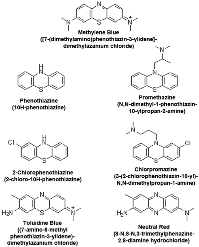
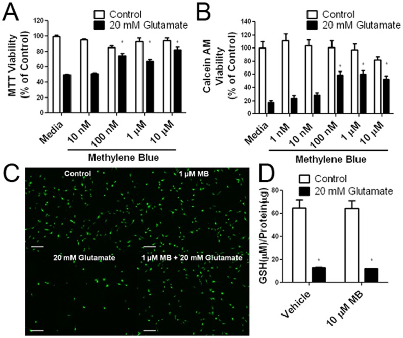
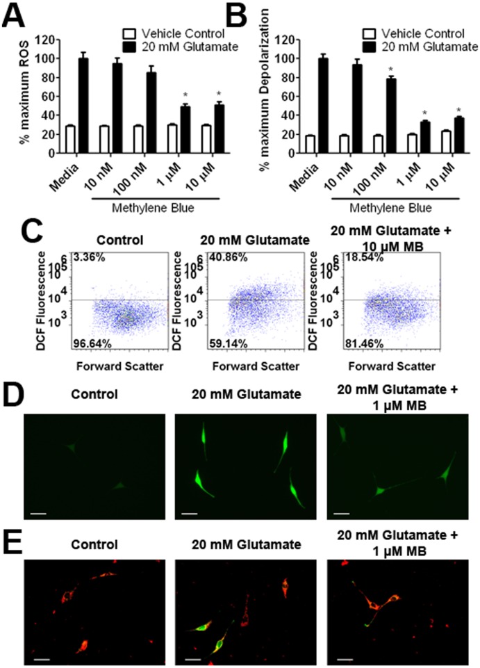
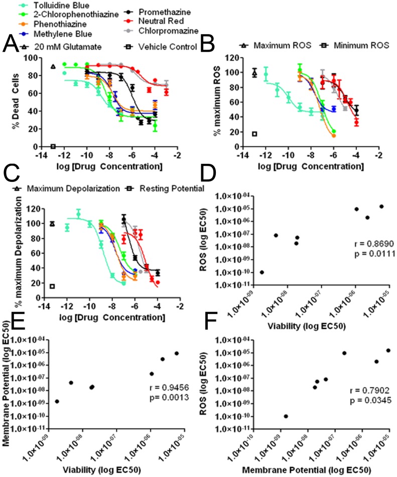
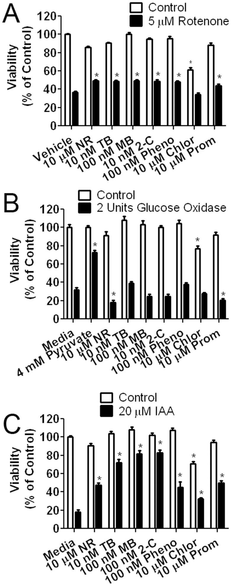
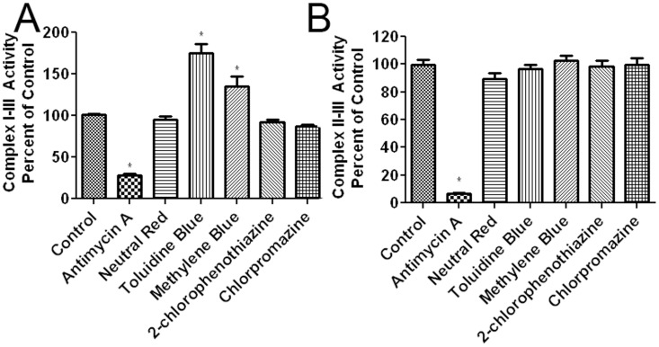
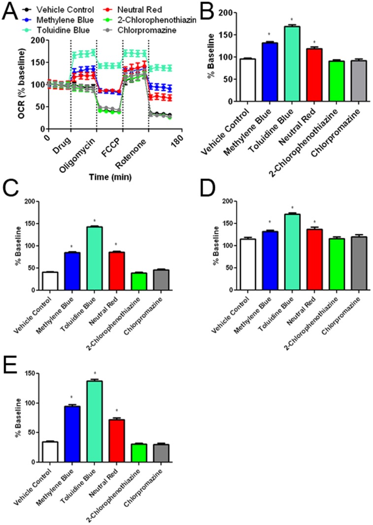
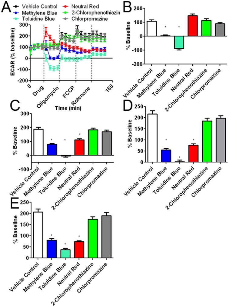
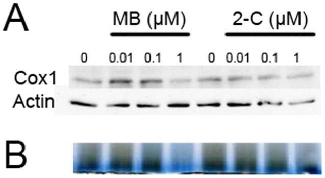
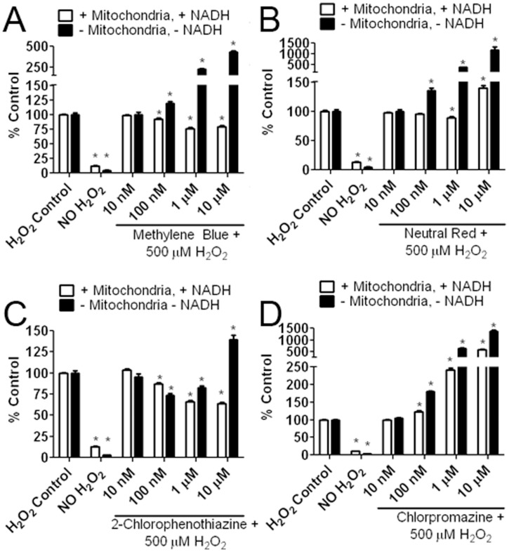
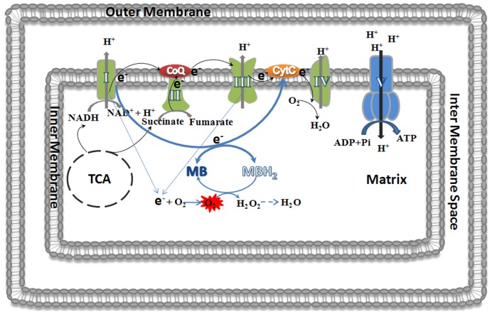
Methylene blue (MB), the first lead chemical structure of phenothiazine and other derivatives, is commonly used in diagnostic procedures and as a treatment for methemoglobinemia. We have previously demonstrated that MB could function as an alternative mitochondrial electron transfer carrier, enhance cellular oxygen consumption, and provide protection in vitro and in rodent models of Parkinson's disease and stroke. In the present study, we investigated the structure-activity relationships of MB in vitro using MB and six structurally related compounds. MB reduces mitochondrial superoxide production via alternative electron transfer that bypasses mitochondrial complexes I-III. MB mitigates reactive free radical production and provides neuroprotection in HT-22 cells against glutamate, IAA and rotenone toxicity. Distinctly, MB provides no protection against direct oxidative stress induced by glucose oxidase. Substitution of a side chain at MB's 10-nitrogen rendered a 1000-fold reduction of the protective potency against glutamate neurototoxicity. Compounds without side chains at positions 3 and 7, chlorophenothiazine and phenothiazine, have distinct redox potentials compared to MB and are incapable of enhancing mitochondrial electron transfer, while obtaining direct antioxidant actions against glutamate, IAA, and rotenone insults. Chlorophenothiazine exhibited direct antioxidant actions in mitochondria lysate assay compared to MB, which required reduction by NADH and mitochondria. MB increased complex IV expression and activity, while 2-chlorphenothiazine had no effect. Our study indicated that MB could attenuate superoxide production by functioning as an alternative mitochondrial electron transfer carrier and as a regenerable anti-oxidant in mitochondria.


Conflict of interest statement

Competing Interests: The authors have declared that no competing interests exist.

Figures

Figure 1
Figure 1. Chemical Structure and IUPAC designation of MB and its related compounds.
Figure 2
Figure 2. Neuroprotective effect of MB against glutamate neurotoxicity in HT-22 cells.
(A) MTT viability assay (B) and Calcein AM viability assay at 12 hours after 20 mM glutamate insult demonstrate the dose dependent neuroprotective effect of MB. * p<0.05 compared to 20 mM glutamate media (C) Representative Calcein AM fluorescent images depict the neuroprotective effect of MB (1 µM) in HT-22 cells after 12 hours exposure of 20 mM glutamate (scale bar 200 µm). (D) Total GSH assay demonstrates that MB has no effect on the glutamate-induced GSH depletion. * p<0.05 compared to vehicle control.
Figure 3
Figure 3. Effect of MB on ROS production and mitochondrial membrane potential depolarization induced by glutamate in HT-22 cells.
(A) DCF microplate reader assay depicts that a significant increase of ROS was induced by a 12 hour exposure to 20 mM glutamate, which was dose dependently attenuated MB. (B) TMRE/NAO plate reader assay depicts a significant mitochondria membrane potential depolarization induced by glutamate insult, which was dose dependently attenuated by MB. (C) Representative DCF flow cytometry assay depicts increase of ROS induced by 8 hour exposure of 10 mM glutamate which was attenuated by 10 µM MB. (D) Representative images of DCF fluorescence demonstrated increased cellular ROS after 8 hours exposure to 20 mM glutamate. DCF fluorescence was reduced with co-treatment of 1 µM MB (scale bar 50 µm). (E) Representative images of JC-1 fluorescence indicate mitochondria membrane potential collapse after an 8 hour exposure to 20 mM glutamate, which was attenuated by co-treatment of 1 µM MB (scale bar 50 µm). * p<0.05 compared to 20 mM glutamate media.
Figure 4
Figure 4. Dose response curves of MB and derivatives in the HT-22 glutamate model.
(A) Dose response curves of MB and derivative against glutamate-induced neurotoxicity measured by Calcein AM; (B) Dose response curves of MB and derivatives against glutamate-induced cellular ROS production measured by DCF assay; (C) Dose response curves of MB and derivatives against mitochondria membrane potential depolarization induced by glutamate measured by NAO/TMRE FRET assay; (D) Correlation of cellular ROS production and cell viability, Pearson coefficient  = 0.8690, p  = 0.00111; (E) Correlation of mitochondria membrane potential and cell viability, Pearson coefficient  = 0.9456, p  = 0.0013; (F) Correlation of cellular ROS production and mitochondria membrane potential, Pearson coefficient  = 0.7902, p  = 0.0345.
Figure 5
Figure 5. Effect of MB and its derivatives in rotenone, IAA, and glucose oxidase toxicity assays.
(A) Effect of MB and derivatives against rotenone neurotoxicity in HT-22 cells. Cells were exposed to 5 µM rotenone for 24 hours in the presence of MB or its derivatives. Protective effect was observed in all compounds except chlorpromazine at the indicated concentrations (NR – Neutral Red; TB – Toluidine Blue O; MB – Methylene Blue; 2-C –2-chlorophenothiazine; Pheno – Phenothiazine; Chlor – Chlorpromazine Prom – Promethazine); # p<0.05 compared to media control. * p<0.05 compared to 5 µM rotenone in media (B) Effect of MB and derivatives against glucose oxidase neurotoxicity in HT-22 cells. Cells were exposed to 2 U glucose oxidase for 3 hours in the presence of MB or derivatives. No protective effect was observed in all compounds tested. Pyruvate (4 mM) was used as a positive control. # p<0.05 compared to media control. * p<0.05 compared to 5 U Glucose Oxidase in media (C) Effect of MB and derivatives against IAA neurotoxicity in HT-22 cells. Cells were exposed to 20 µM IAA for 24 hours in the presence of MB or derivatives. Protective effect was observed in all compounds at the indicated concentration. # p<0.05 compared to media control. * p<0.05 compared to 20 µM IAA in media.
Figure 6
Figure 6. Effect of MB and derivatives on mitochondrial complexes activity.
(A) The effects of MB and its derivatives on the rate of cytochrome c reduction were recorded by measuring the change in absorbance of reduced cytochrome c with NADH as an electron donor (complex I-III activity) in isolated mitochondria. All compounds were tested at the concentration of 10 µM. MB and TB significantly increased complex I-III activity (the rate of cytochrome c reduction). Antimycin A (2 µg/ml), a complex III inhibitor, was used as a negative control. (B) Mitochondria complex II-III was measured by the change in absorbance of reduced cytochrome c with succinate as an electron donor in isolated mitochondria. All compounds were tested at the concentration of 10 µM. No effect was observed in all MB related compounds. Antimycin A (2 µg/ml) significantly decreased the rate of cytochrome c reduction. * p<0.05 compared to control.
Figure 7
Figure 7. Effects of the MB and its derivatives on cellular oxygen consumption rate (OCR).
(A) OCR recording at baseline and subsequent treatment of MB or its derivatives, oligomycin, FCCP, and rotenone. The initial 35 minutes establishes a baseline reading, followed by addition of each drug at a concentration of 10 µM. Three subsequent injections followed consisting of 1 µg/mL oligomycin (complex V inhibitor), 300 nM FCCP (proton gradient uncoupler), and 100 nM rotenone (complex I inhibitor). After each injection, 4 time points were recorded with about 35 minutes between each injection. (B) MB, TB, and NR increased oxygen consumption as compared to vehicle control. 2-Chlorophenothiazine and chlorpromazine had no effect compared to vehicle. (C) Oligomycin decreased cellular oxygen consumption under all experimental conditions. Despite the oligomycin insult, MB, TB and NR significantly increased OCR as compared to vehicle control. (D) Injection of FCCP results in maximum cellular OCR. MB, NR, and TB treated groups have higher maximal respiration than vehicle control. (D) Rotenone inhibits complex I causing a decrease in OCR, which was significantly attenuated by the treatment of MB, NR, and TB. * p<0.05 compared to control group.
Figure 8
Figure 8. Effects of the MB and its derivatives on extracellular acidification rate (ECAR).
(A) ECAR recording at baseline and subsequent addition of MB or its derivatives, oligomycin, FCCP, and rotenone. The initial 35 minutes establishes a baseline reading, followed by addition of drug at a concentration of 10 µM. Three subsequent injections followed consisting of 1 µg/ml oligomycin (complex V inhibitor), 300 nM FCCP (proton gradient uncoupler), and 100 nM rotenone (complex I inhibitor). After each injection, 4 time points were recorded with about 35 minutes between injections. (B) MB and TB decreased ECAR as compared to the vehicle control. 2-Chlorophenothiazine and chlorpromazine had no effect as compared to the control. NR, had no effect to vehicle on average, although a spike in ECAR values was observed after NR injection. (C) Oligomycin increased ECAR under all experimental conditions and was significantly reduced by MB, TB, and NR. (D) Injection of FCCP results in maximum cellular OCR with little effect on ECAR; MB, NR, and TB significantly decreased ECAR as compared to the vehicle control. (D) Rotenone inhibits complex I and decreases OCR without change in ECAR. MB, NR and TB caused ECAR to remain significantly less as compared to the vehicle control. * p<0.05 compared to control group.
Figure 9
Figure 9. Different action of MB and 2-chlorophenothiazine on mitochondrial complex IV.
(A) Western blots depict the expression of complex IV subunit I (Cox1) in HT-22 cells treated with MB or 2-chlorophenothiazine at the indicated concentrations for 3 days. MB, at concentrations of 10 and 100 nM, increased Cox1expression. 2-Chlorophenothiazine had no effect on Cox1 expression at 10 nM, 100 nM, and 1 µM. (B). Blue native indicated an increase in complex IV activity at 100 nM MB and a decrease in complex IV activity at 1 µM MB corresponding to the increased expression of Cox1. 2-Chlorophenothiazine had no effect on complex IV activity at all concentrations tested.
Figure 10
Figure 10. Different action of MB and derivatives as antioxidants.
Four compounds were assayed in the presence or absence of mitochondria lysate and 165 µM NADH to determine their effectiveness in mitigating H2O2 (500 µM) induced DCF oxidation. (A) In the presence of mitochondria lysate and NADH, MB significantly reduced DCF fluorescence at 100 nM, 1 µM and 10 µM. At the same concentrations (100 nM, 1 µM, and 10 µM) in the absence of mitochondria and NADH, MB increased DCF fluorescence. (B) In the presence of mitochondria and NADH, NR decreased DCF fluorescence at a concentration of 1 µM and increased DCF fluorescence at a concentration of 10 µM. NR significantly increased DCF fluorescence at concentrations of 100 nM, 1 µM, and 10 µM in the absence of mitochondria lysate and NADH. (C) 2-Chlorophenothiazine significantly reduced DCF fluorescence at concentrations of 100 and 1 µM in both the presence and absence of mitochondria lysate and NADH. At a concentration of 10 µM, in the presence of mitochondria lysate and NADH, 2-chlorophenothiazine reduced DCF fluorescence; however, in the absence of mitochondria lysate and NADH, 2-chlorophenothiazine increased DCF fluorescence at 10 µM. (D) Chlorpromazine significantly increased DCF fluorescence at concentrations of 100 nM, 1 µM, and 10 µM in both the presence and absence of mitochondria lysate and NADH. * p<0.05 compared to respective H2O2 control group.
Figure 11
Figure 11. Schematic illustration depicts the novel neuroprotective mechanism of MB.
MB receives electrons from NADH through mitochondrial complex I and is reduced to leuco-MB, which can donate the electrons to cytochrome c and recycle to its oxidized form, MB. In addition, the reduced leuco-MB can also function as free radical scavenger and neutralize superoxide generated due to the blockage of complex I and III.

References

    1. Bergen DC (2008) Neurological Disorders: Public Health Challenges. Arch Neurol 65: 154–154.
    1. Pratico D (2008) Oxidative stress hypothesis in Alzheimer’s disease: a reappraisal. Trends Pharmacol Sci 29: 609–615. - PubMed
    1. Barnham KJ, Masters CL, Bush AI (2004) Neurodegenerative diseases and oxidative stress. Nat Rev Drug Discov 3: 205–214. - PubMed
    1. Sanchez-Moreno C, Dashe JF, Scott T, Thaler D, Folstein MF, et al. (2004) Decreased levels of plasma vitamin C and increased concentrations of inflammatory and oxidative stress markers after stroke. Stroke 35: 163–168. - PubMed
    1. Kamat CD, Gadal S, Mhatre M, Williamson KS, Pye QN, et al. (2008) Antioxidants in central nervous system diseases: preclinical promise and translational challenges. J Alzheimers Dis 15: 473–493. - PMC - PubMed

Publication types

MeSH terms