. 2012;7(10):e48608.
doi: 10.1371/journal.pone.0048608. Epub 2012 Oct 31.

C. difficile 630Δerm Spo0A regulates sporulation, but does not contribute to toxin production, by direct high-affinity binding to target DNA

Affiliations

C. difficile 630Δerm Spo0A regulates sporulation, but does not contribute to toxin production, by direct high-affinity binding to target DNA

Katharina E Rosenbusch et al. PLoS One. 2012.

Abstract

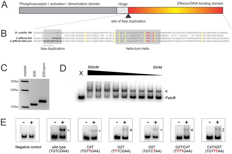
Clostridium difficile is a Gram positive, anaerobic bacterium that can form highly resistant endospores. The bacterium is the causative agent of C. difficile infection (CDI), for which the symptoms can range from a mild diarrhea to potentially fatal pseudomembranous colitis and toxic megacolon. Endospore formation in Firmicutes, including C. difficile, is governed by the key regulator for sporulation, Spo0A. In Bacillus subtilis, this transcription factor is also directly or indirectly involved in various other cellular processes. Here, we report that C. difficile Spo0A shows a high degree of similarity to the well characterized B. subtilis protein and recognizes a similar binding sequence. We find that the laboratory strain C. difficile 630Δerm contains an 18bp-duplication near the DNA-binding domain compared to its ancestral strain 630. In vitro binding assays using purified C-terminal DNA binding domain of the C. difficile Spo0A protein demonstrate direct binding to DNA upstream of spo0A and sigH, early sporulation genes and several other putative targets. In vitro binding assays suggest that the gene encoding the major clostridial toxin TcdB may be a direct target of Spo0A, but supernatant derived from a spo0A negative strain was no less toxic towards Vero cells than that obtained from a wild type strain, in contrast to previous reports. These results identify for the first time direct (putative) targets of the Spo0A protein in C. difficile and make a positive effect of Spo0A on production of the large clostridial toxins unlikely.


Conflict of interest statement

Competing Interests: The authors have declared that no competing interests exist.

Figures

Figure 1
Figure 1. Purification and detection of C. difficile Spo0A. A.
Heterologous overproduction of Spo0A-6xHis and Spo0A-DBD-6xHis in E. coli Rosetta(DE3) pLysS. M  =  molecular weight marker, numbers indicate hours after induction with 1 mM IPTG. P  =  metal affinity purified protein. Lysates were separated on a 12% SDS-PAGE. B and C. Immunoblot detection of Spo0A in total cell lysates of a C. difficile spo0A mutant (CT::spo0A) and a wild type strain (630Δerm). Times indicated range from early exponential (3 h post inoculation) to late stationary growth phase (48 h post inoculation). Sample volumes were corrected for OD600 to ensure loading of similar amounts of total cell lysate in each lane. For details see Materials and Methods. M  =  molecular weight marker. B. ECL+ detection. C. Fluorescent detection. Y-axes show peak volumes normalized to values at 48 hours post inoculation (closed diamonds; left axis) and optical density readings at 600 nM (open squares; right axis). Inset shows the blot on which the curve is based. Vertical dashed line indicates the moment Spo0A levels increase sharply (6 hours post inoculation).
Figure 2
Figure 2. A Spo0A box is important for high affinity binding by C. difficile Spo0A. A.
Domain organization of Spo0A. The site of the duplication in strain 630Δerm identified in this study is indicated by an arrow. B. Sequence alignment of the C-terminal regions of the Spo0A proteins of B. subtilis 168 and C. difficile strains 630 and 630Δerm. Residues identified in structural studies using Bacillus Spo0A as involved in backbone interactions are indicated in yellow, residues forming base-specific contacts are indicated in red . The region of the 6aa duplication and the helix-turn-helix motifs are boxed in gray and the duplication in strain 630Δerm is underlined. C. PCR showing the presence of the duplication near the DNA binding domain in C difficile 630Δerm compared to 630. D. Electrophoretic mobility shift assay using purified C. difficile Spo0A-DBD-6xHis and a radiolabeled PabrB DNA fragment. X  =  no protein control, the triangle indicates 1.3-fold serial dilutions of protein to the indicated concentrations. The arrow indicates a DNA:protein complex. E. Electrophoretic mobility shift assays without (−) or with (+) 150 nM Spo0A-DBD-6xHis added to radiolabeled PabrB fragments carrying mutations in the consensus Spo0A box (in red). Arrows indicate DNA:protein complexes. The negative control is PcitG from B. subtilis.
Figure 3
Figure 3. C. difficile Spo0A binds to predicted and expected target sequences.
Electrophoretic mobility shift assay using purified C. difficile Spo0A-DBD-6xHis and a radiolabeled DNA fragments. X  =  no protein control, the triangle indicates 1.3-fold serial dilutions of protein to the indicated concentrations. The arrows indicate DNA:protein complexes. A. In silico predicted target sequences upstream of the genes encoding Spo0A (spo0A), σH (sigH), a lipoate ligase (lplA) and an aliphatic sulphonates ABC transporter (ssuA). B. Target sequences predicted on the basis of findings in other organisms: spoIIAA, spoIIE and spoIIGA. The DNA upstream of spoVG serves as a negative control.
Figure 4
Figure 4. C. difficile Spo0A does not contribute to toxin production directly. A.
Electrophoretic mobility shift assays using purified C. difficile Spo0A-DBD-his6 and radiolabeled DNA fragments corresponding to the upstream regions of the indicated toxin-related genes. X  =  no protein control, the triangle indicates 1.3-fold serial dilutions of protein to the indicated concentrations. Arrows indicate (possible) protein:DNA complexes. B. Toxin end-point titre of culture supernatant from wild type (630Δerm; white bars) or spo0A mutant (CT::spo0A; black bars) C. difficile cells. For details see Materials and Methods. Error bars represent standard error of the mean (n = 3). None of the observed differences were significant in an independent sample t-test.

References

    1. Errington J (2003) Regulation of endospore formation in Bacillus subtilis . Nat Rev Microbiol 1: 117–126. 10.1038/nrmicro750 [doi]. - PubMed
    1. Paredes CJ, Alsaker KV, Papoutsakis ET (2005) A comparative genomic view of clostridial sporulation and physiology. Nat Rev Microbiol 3: 969–978. nrmicro1288 [pii];10.1038/nrmicro1288 [doi]. - PubMed
    1. Ferrari FA, Trach K, LeCoq D, Spence J, Ferrari E, et al. (1985) Characterization of the spo0A locus and its deduced product. Proc Natl Acad Sci U S A 82: 2647–2651. - PMC - PubMed
    1. Bird TH, Grimsley JK, Hoch JA, Spiegelman GB (1993) Phosphorylation of Spo0A activates its stimulation of in vitro transcription from the Bacillus subtilis spoIIG operon. Mol Microbiol 9: 741–749. - PubMed
    1. Baldus JM, Green BD, Youngman P, Moran CP Jr (1994) Phosphorylation of Bacillus subtilis transcription factor Spo0A stimulates transcription from the spoIIG promoter by enhancing binding to weak 0A boxes. J Bacteriol 176: 296–306. - PMC - PubMed

Publication types

MeSH terms

Associated data

LinkOut - more resources