Review
. 2013:64:307-24.
doi: 10.1146/annurev-med-072211-110606. Epub 2012 Nov 1.

Defective complement inhibitory function predisposes to renal disease

Affiliations
Review

Defective complement inhibitory function predisposes to renal disease

Anuja Java et al. Annu Rev Med. 2013.

Abstract

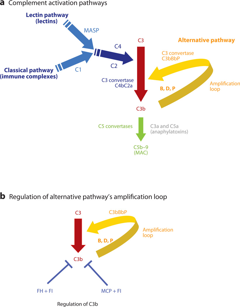
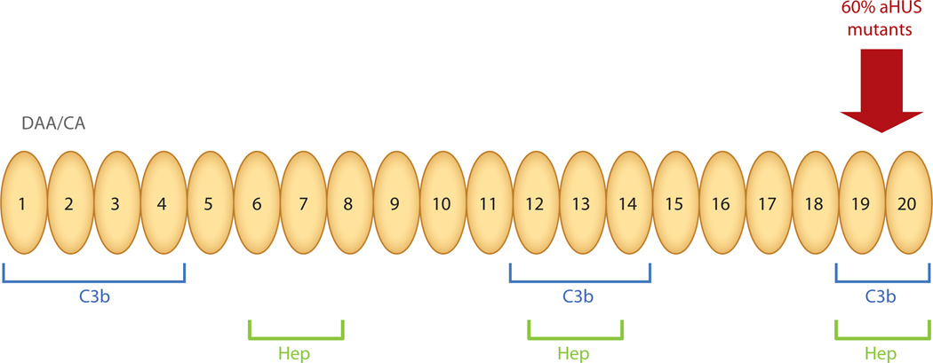
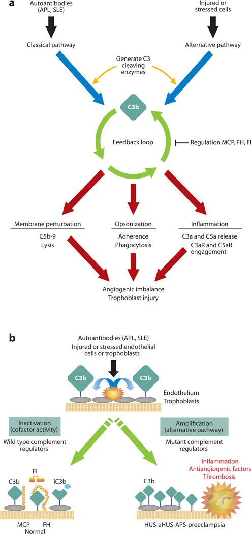
The role of the complement system in mediating human renal disease has long been recognized in immune-complex excess syndromes such as systemic lupus erythematosus and in dense deposit disease in which no immunoglobulin (Ig) is present. Over the past 15 years, mutations in complement regulatory genes have been demonstrated to predispose to thrombotic microangiopathies including atypical hemolytic uremic syndrome, C3 and C1q glomerulopathies, and preeclampsia. Excessive complement activation on an endothelial cell, due to either an autoantibody or a regulatory protein deficiency, sets up a procoagulant state in these diseases as well as in the antiphospholipid syndrome. Knowledge of the genes involved and the functional consequences of alterations in their structure has led to therapy that blocks complement activation.


Figures

Figure 1
Figure 1
Complement activation pathways. (a) Activation of the classical, lectin, and alternative pathways results in the generation of C3 convertases that cleave C3 into C3b and C3a. C1 of the classical pathway and mannose-binding associated serine proteases (MASPs) of the lectin pathway cleave C4 and C2, and the resultant larger fragments then form the classical/lectin pathway C3 convertase. C3 is cleaved by C3 convertases into C3b, which deposits on the cell surface, and C3a, an anaphylatoxin, which recruits and activates effector cells. C3b may rapidly amplify through a positive feedback loop termed the C3b alternative pathway amplification loop. C3b may also trigger activation of C5 through the generation of the C5 convertases, which are composed of C3b attached to a C3 convertase. This structure alters the substrate specificity of the convertase to favor C5. C5 convertase cleaves C5 and results in the generation of C5a (an anaphylatoxin) and assembly of the membrane attack complex (MAC) beginning with C5b (plus C6, C7, C8 and many C9s). (b) The amplification or feedback loop of the alternative pathway. Factor H (FH), factor I (FI), and membrane cofactor protein (MCP) are the key regulators of the alternative pathway and interrupt the feedback loop. Excessive activation via this loop secondary to a decrease in regulatory activity or a gain of function in a component like FB or C3 predisposes to atypical hemolytic uremic syndrome. Also see Table 1. FI is a serine protease; in conjunction with a cofactor protein (FH or MCP), it cleaves C3b to iC3b, which cannot form a C3 convertase, i.e., the feedback loop cannot be engaged. C3bBbP is the alternative-pathway C3 convertase.
Figure 2
Figure 2
Schematic diagram of factor H. The N-terminal repeats form the regulatory domain and the C-terminal repeats 19–20 form the major surface recognition domain. The majority of mutations reported in aHUS are in the COOH terminal repeats 19–20. Weaker C3b binding and recognition domains are also located in repeats 6–7 and 12–14. The surface-binding repeats are also known as anionic or heparin-binding sites. DAA, decay-accelerating activity; CA, cofactor activity; Hep, heparin binding. Adapted from Reference with permission.
Figure 3
Figure 3
Complement activation on injured or stressed cells. (a) Black and blue arrows: autoantibodies (such as antiphospholipid antibodies and other SLE-associated antibodies) and damaged tissues trigger the complement cascade; green arrows: deposition of C3b on a target sets in motion the powerful amplification loop of the alternative pathway; red arrows: effector activities of complement are generated by C3b deposition and C3a release and the downstream mediators C5b–9 and C5a. Reprinted from Reference . (b) Regulation of feedback loop of alternative pathway on injured or stressed cells. This occurs through limited proteolytic cleavage of C3b to generate iC3b. The reaction is carried out by a serine protease factor I (FI) and membrane cofactor protein (MCP) or factor H (FH). Cofactor activity terminates the feedback loop because iC3b does not bind FB, thereby shutting down the amplification loop. Because APL are autoantibodies that trigger the classical pathway, defective regulation of C4b (a component of the classical pathway C3 convertase) by MCP is also likely to influence the severity of tissue injury and risk for preeclampsia. When tissues are damaged, a delicate balance must be established to allow for repair and recovery. If regulators such as MCP, FI, or FH are dysfunctional, excessive complement activation occurs. APS, antiphospholipid syndrome; SLE, systemic lupus erythematosus. Adapted from Reference with permission.

References

    1. Markiewski MM, Nilsson B, Ekdahl KN, et al. Complement and coagulation: strangers or partners in crime? Trends Immunol. 2007;28:184–192. - PubMed
    1. Pickering MC, Cook HT. Translational mini-review series on complement factor H: renal diseases associated with complement factor H: novel insights from humans and animals. Clin. Exp. Immunol. 2008;151:210–230. - PMC - PubMed
    1. Richards A, Kavanagh D, Atkinson JP. Inherited complement regulatory protein deficiency predisposes to human disease in acute injury and chronic inflammatory states: the examples of vascular damage in atypical hemolytic uremic syndrome and debris accumulation in age-related macular degeneration. Adv. Immunol. 2007;96:141–177. Recent, comprehensive, and authoritative reviews on aHUS.

    1. Abarrategui-Garrido C, Martinez-Barricarte R, Lopez-Trascasa M, et al. Characterization of complement factor H-related (CFHR) proteins in plasma reveals novel genetic variations ofCFHR1 associated with atypical hemolytic uremic syndrome. Blood. 2009;114:4261–4271. - PubMed
    1. Atkinson JP, Goodship TH. Complement factor H and the hemolytic uremic syndrome. J. Exp. Med. 2007;204:1245–1248. - PMC - PubMed

MeSH terms