. 2013 May 1;18(13):1549-56.
doi: 10.1089/ars.2012.5037. Epub 2012 Dec 10.

Is oxidized thioredoxin a major trigger for cysteine oxidation? Clues from a redox proteomics approach

Affiliations

Is oxidized thioredoxin a major trigger for cysteine oxidation? Clues from a redox proteomics approach

Sarela García-Santamarina et al. Antioxid Redox Signal. .

Abstract

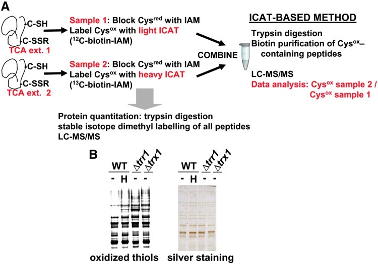
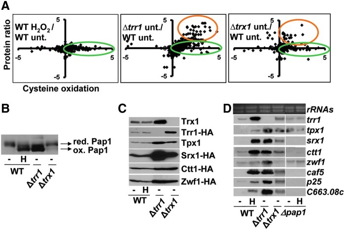
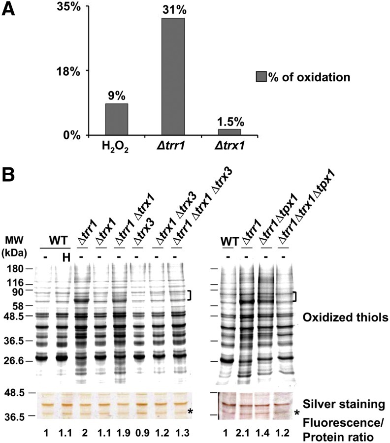
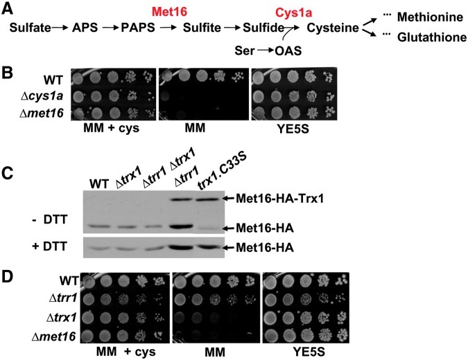
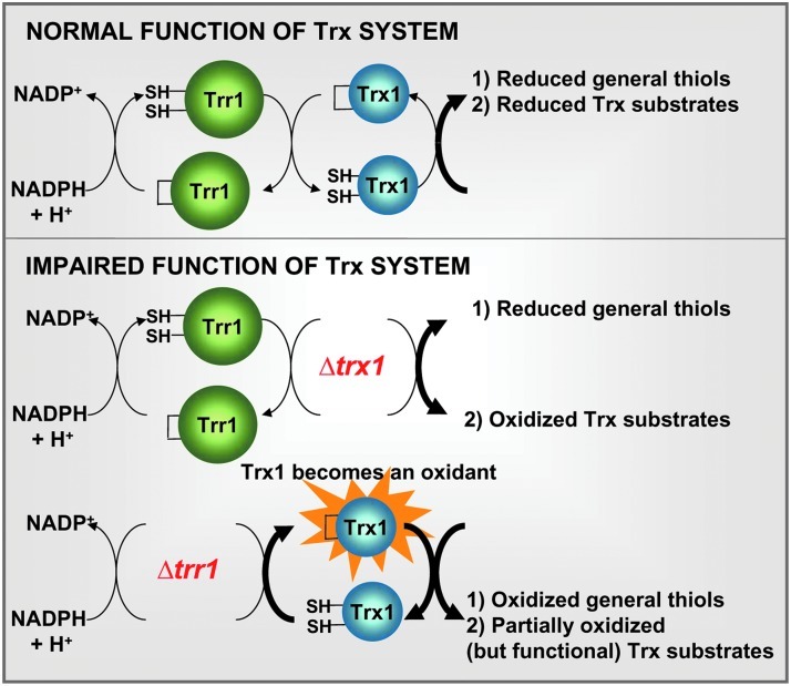
Cysteine oxidation mediates oxidative stress toxicity and signaling. It has been long proposed that the thioredoxin (Trx) system, which consists of Trx and thioredoxin reductase (Trr), is not only involved in recycling classical Trx substrates, such as ribonucleotide reductase, but it also regulates general cytoplasmic thiol homeostasis. To investigate such a role, we have performed a proteome-wide analysis of cells expressing or not the two components of the Trx system. We have compared the reversibly oxidized thiol proteomes of wild-type Schizosaccharomyces pombe cells with mutants lacking Trx or Trr. Specific Trx substrates are reversibly-oxidized in both strain backgrounds; however, in the absence of Trr, Trx can weakly recycle its substrates at the expense of an alternative electron donor. A massive thiol oxidation occurs only in cells lacking Trr, with 30% of all cysteine-containing peptides being reversibly oxidized; this oxidized cysteine proteome depends on the presence of Trxs. Our observations lead to the hypothesis that, in the absence of its reductase, the natural electron donor Trx becomes a powerful oxidant and triggers general thiol oxidation.


Figures

FIG. 1.
FIG. 1.
Isotope-coded affinity tag (ICAT) strategy for studying the in vivo status of reversibly oxidized cysteines in different strain backgrounds and conditions. (A) Schematic representation of the ICAT methodology. Trichloroacetic acid (TCA) protein extracts were obtained for each pair of samples to be analyzed at a time. Thiols (Cysred) in the extracts were alkylated with iodoacetamide (IAM). Upon reduction of oxidized thiols (Cysox), resulting thiols were alkylated with either light (12C-biotin-IAM) or heavy (13C-biotin-IAM) ICAT reagent. Labeled protein extracts were then mixed and digested with trypsin. ICAT-labeled peptides were affinity purified through streptavidin columns, fractionated by liquid chromatography, and analyzed by mass spectrometry (LC-MS/MS). To quantify individual proteins by dimethyl labeling, small fractions of protein extracts were digested with trypsin, and resulting peptides were labeled at their amino groups with light or heavy formaldehyde (dimethyl labeling). Resulting peptides were mixed and fractionated by LC-MS/MS. (B) Labeling of reversibly oxidized cysteines for 1D electrophoresis. Free thiols in TCA protein extracts of untreated (−) or treated (0.2 mM H2O2 for 30 s; H) cultures of strains 972 (WT), SG167 (Δtrr1), and MJ15 (Δtrx1) were alkylated with iodoacetamide. Upon reduction of oxidized thiols, resulting thiols were alkylated with a fluorescently labeled iodoacetamide derivative. Samples were analyzed by fluorescent 1D gel electrophoresis (oxidized thiols) and with silver staining, as a control of protein loading. (To see this illustration in color, the reader is referred to the web version of this article at www.liebertpub.com/ars.)
FIG. 2.
FIG. 2.
Over-expression of proteins in Δtrr1 and in Δtrx1 cells is partially dependent on the transcription factor Pap1. (A) ICAT data representation: cysteine oxidation versus protein expression in the three ICAT studied pairs. Cysteine oxidation from four experimental conditions was analyzed pairwise using the ICAT method depicted in Figure 1A. In each panel, the Log2 ratio of cysteine oxidation is plotted in a scatter diagram versus its Log2 protein ratio (72%–78% of the cysteine-containing peptides displayed protein values by dimethyl labeling). Left panel: 972 treated with 0.2 mM H2O2 for 30 s versus 972 untreated (WT H2O2/WT unt.). Center panel: NG25 untreated versus 972 untreated (Δtrr1 unt./WT unt.). Right panel: MJ15 untreated versus 972 untreated (Δtrx1 unt./WT unt.). Green circles represent peptides with increased cysteine oxidation and orange circles represent peptides from over expressed proteins. (B–D) The activity of the transcription factor Pap1 explains increased protein levels in some strain backgrounds. (B) In vivo oxidation of Pap1. Cultures of strains IC2 (WT), IC71 (Δtrr1), and MJ2 (Δtrx1) were treated (H) or not (−) with 0.2 mM H2O2 for 5 min. TCA extracts were obtained and analyzed by nonreducing electrophoresis. Reduced/inactive (red.) and oxidized/active (ox.) Pap1 forms are indicated with arrows. (C) Protein levels of some Pap1-dependent targets. Cultures of strains expressing or not tagged proteins were treated (H) or not (−) with 0.2 mM H2O2 for 5 min. TCA extracts were obtained, and specific Pap1-dependent proteins were analyzed from extracts of strains: 972, SG167 (Δtrr1) and MJ15 (Δtrx1) for Trx1 and Tpx1; MJ8 (trr1-HA WT), SG167 (Δtrr1), and SG202 (trr1-HA Δtrx1) for Trr1-HA; SB50 (srx1-HA WT), SB69 (srx1-HA Δtrr1), and SB68 (srx1-HA Δtrx1) for Srx1-HA; JF17 (ctt1-HA WT), SG200 (ctt1-HA Δtrr1), and SG198 (ctt1-HA Δtrx1) for Ctt1-HA; and SG18 (zwf1-HA WT), in SB32 (zwf1-HA Δtrr1), and in SG37 (zwf1-HA Δtrx1) for Zwf1-HA. Western blots were performed using polyclonal anti-Tpx1 or anti-Trx1 antibodies, or monoclonal anti-HA antibodies. (D) Transcriptional analysis of Pap1-dependent genes. RNA from strains 972 (WT), IC1 (Δpap1), IC71 (Δtrr1), and MJ2 (Δtrx1), untreated (−) or treated with 0.2 mM H2O2 for 15 min (H), was obtained and analyzed by Northern blot with probes for trr1, tpx1, srx1, ctt1, zwf1, caf5, p25, and SPCC663.08c. Ribosomal RNA (rRNAs) was used as a loading control.
FIG. 3.
FIG. 3.
General oxidation of thiols in Δtrr1 cells is dependent on the presence of oxidized cytoplasmic Trx1 and Trx3 and/or Tpx1. (A) Percentage of oxidized cysteines in the different strains according to ICAT data. For each peptide in each biological condition (wild-type treated with H2O2; untreated Δtrr1; or untreated Δtrx1) a ratio of oxidation was always obtained comparing to untreated wild-type cells (WT unt.). An oxidation average ratio was calculated for those peptides having values>1.5-fold in 2 out of 3 biological replicates (for wild-type treated with H2O2 and untreated Δtrr1 samples), or>1.5-fold in 2 out of 2 biological replicates (for Δtrx1 samples). For those peptides having values of protein quantification by dimethyl labeling, a ratio was calculated as oxidation average ratio/protein levels, and only those having this ratio>1.5-fold are included in this graph. For peptides not displaying values on protein concentration, we eliminated those regulated by Pap1, and those having an oxidation average ratio>1.5-fold are included in this graph. Bars represent the percentage of cysteine-containing peptides, which fulfill the previous criteria for each experimental condition. (B) Role of the Trx/Trr system in the homeostasis of cysteine protein oxidation. Free thiols from TCA protein extracts from untreated (−) or treated (0.2 mM H2O2, 30 s; H) strains 972 (WT), SG167 (Δtrr1), MJ15 (Δtrx1), PG22 (Δtrr1 Δtrx1), SG189 (Δtrx3), IC76 (Δtrx1 Δtrx3), SG185 (Δtrr1 Δtrx1 Δtrx3), SG164 (Δtrr1 Δtpx1), and SG170 (Δtrr1 Δtrx1 Δtpx1) were processed as described in Figure 1B. The intensity of selected fluorescent labeled proteins, indicated in the figure with left braces, was quantified with ImageQuant. The intensity of selected proteins of the silver staining gel, indicated with an asterisk, was quantified with ImageJ. Fluorescence to protein ratios were calculated and are indicated in the figure. (To see this illustration in color, the reader is referred to the web version of this article at www.liebertpub.com/ars.)
FIG. 4.
FIG. 4.
In the absence of its reductase, Trx1 is able to recycle substrates at the expense of an alternative electron donor. (A) Cysteine biosynthesis pathway in Schizosaccharomyces pombe. The enzymatic roles of Met16 and Cys1a are indicated. (B) Exponentially growing 972 (WT), SG178 (Δcys1a), and SG171 (Δmet16) strains were serially diluted and spotted on minimal medium (MM), MM containing 0.66 mM cysteine (MM+cys), and YE5S plates. (C) Mixed disulfide of Met16 with Trx1. Immunodetection of Met16-HA from TCA extracts of strains SG54 (WT), SG71 (Δtrx1), SG78 (Δtrr1 Δtrx1), SG59 (Δtrr1), and SG181 (trx1.C33S), under nonreducing (−DTT, upper panel) and reducing (+DTT, lower panel) electrophoresis. Since cysteine 33 in Trx1 resolves the mixed disulfides with its substrates, the Trx1.C33S mutant allows in vivo trapping of an intermolecular disulfide of Trx1 with Met16. Arrows indicate Met16-HA and Met16-HA covalently linked to Trx1. (D) S. pombe cells lacking Trx1 are auxotrophic for cysteine, whereas S. pombe cells lacking Trr1 are not. Exponentially growing cultures of strains 972 (WT), SG167 (Δtrr1), MJ16 (Δtrx1), and SG171 (Δmet16) were serially diluted and spotted on MM, MM containing 0.66 mM cysteine (MM+cys), and YE5S plates. (To see this illustration in color, the reader is referred to the web version of this article at www.liebertpub.com/ars.)
FIG. 5.
FIG. 5.
Proposed model for the participation of the thioredoxin system in general thiol homeostasis and in the recycling of specific Trx substrates. Thioredoxins specifically recycle proteins that form disulfides as part of their catalytic activities (upper panel). In the absence of Trx (lower panel, Δtrx1), Trx substrates are not recycled and appear as oxidized, but other thiols in proteins remain reduced. Only when Trr1 is absent (lower panel, Δtrr1), thioredoxins are converted into potent oxidants leading to massive reversible thiol oxidation; Trx substrates are partially recycled in this background (see text for details). (To see this illustration in color, the reader is referred to the web version of this article at www.liebertpub.com/ars.)

References

    1. Bulleid NJ. Ellgaard L. Multiple ways to make disulfides. Trends Biochem Sci. 2011;36:485–492. - PubMed
    1. Castillo EA. Ayte J. Chiva C. Moldon A. Carrascal M. Abian J. Jones N. Hidalgo E. Diethylmaleate activates the transcription factor Pap1 by covalent modification of critical cysteine residues. Mol Microbiol. 2002;45:243–254. - PubMed
    1. Chen D. Wilkinson CR. Watt S. Penkett CJ. Toone WM. Jones N. Bahler J. Multiple pathways differentially regulate global oxidative stress responses in fission yeast. Mol Biol Cell. 2008;19:308–317. - PMC - PubMed
    1. Chiappetta G. Ndiaye S. Igbaria A. Kumar C. Vinh J. Toledano MB. Proteome screens for Cys residues oxidation: the redoxome. Methods Enzymol. 2010;473:199–216. - PubMed
    1. Depuydt M. Messens J. Collet JF. How proteins form disulfide bonds. Antioxid Redox Signal. 2011;15:49–66. - PubMed

Publication types