. 2012 Nov 2:7:151.
doi: 10.1186/1746-1596-7-151.

Primary bladder adenocarcinoma versus metastatic colorectal adenocarcinoma: a persisting diagnostic challenge

Affiliations

Primary bladder adenocarcinoma versus metastatic colorectal adenocarcinoma: a persisting diagnostic challenge

Somak Roy et al. Diagn Pathol. .

Abstract

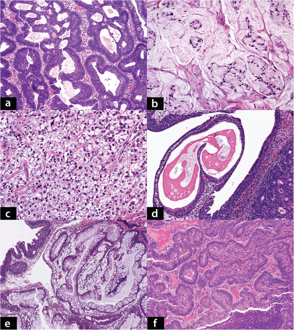
Aim: This study attempted to distinguish primary bladder adenocarcinoma (PBA) from metastatic colonic adenocarcinomas (MCA), which is a difficult diagnostic and clinical problem.

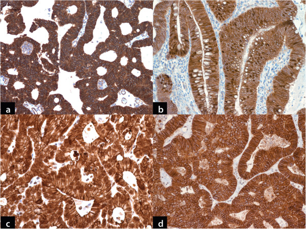
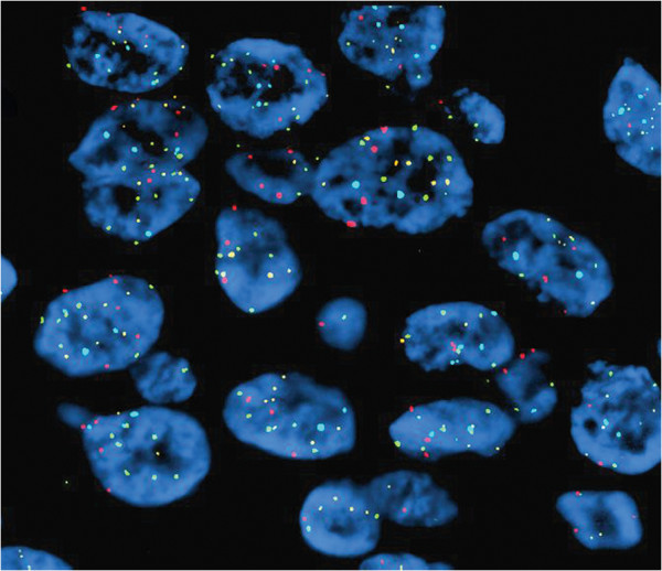
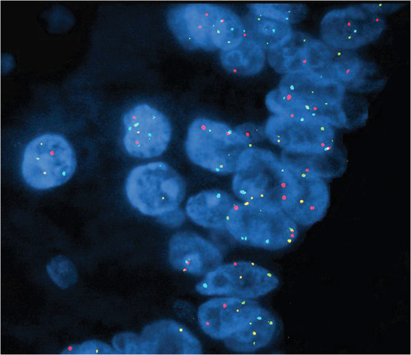
Methods: Twenty-four cases of bladder adenocarcinomas (12 primary & 12 metastatic colorectal) were included in the study with urothelial carcinoma (UC) and colonic adenocarcinoma (CA) as controls. A panel of immunohistochemical (IHC) stains along with fluorescence in-situ hybridization (FISH), using the UroVysion probe set, was performed.

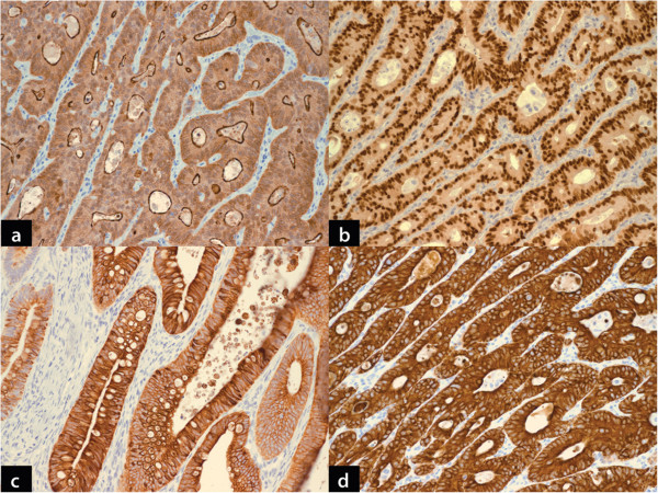
Results: The majority of the PBAs presented with advanced disease. Enteric histologic subtype was the most common morphological variant. Strong nuclear with cytoplasmic-membranous staining of β-catenin was seen in 75% of MCA and only 16.7% PBA (<10% staining cells). Although abnormal nuclear staining with E-cadherin was seen in both PBA and MCA, it was more frequent in former. CK-7, CK-20, villin and CDX-2 stains were not helpful in distinguishing the two entities. FISH did not reveal any unique differences in chromosomal abnormality between the two groups.

Conclusion: Although there was a statistically significant difference in β-catenin and E-cadherin staining between two groups, we did not find any IHC or FISH marker that was specific for PBA. Distinction between PBA and MCA remains a diagnostic problem and clinical correlation is vital before rendering a diagnosis.

Virtual slides: The virtual slides for this article can be found here: http://www.diagnosticpathology.diagnomx.eu/vs/1393156268152357.


Figures

Figure 1
Figure 1
a: Primary bladder adenocarcinoma, enteric type. Moderately differentiated malignant glands are seen with dirty luminal necrosis. Elongated, enlarged and hyperchromatic nuclei with prominent stratification line the malignant glands. (Hematoxylin & eosin, x100). b: Primary bladder adenocarcinoma, mucinous type. Scattered small groups of tumor cells with intracytoplasmic mucin are seen in a background of abundant mucinous material (Hematoxylin & eosin, x200). c: Primary bladder adenocarcinoma, signet ring cell type: Tumor comprised of diffuse sheets of signet ring cells infiltrating the bladder wall (Hematoxylin & eosin, x200). d: Cystitis cystica involving the surface urothelium with underlying invasive primary bladder adenocarcinoma (Hematoxylin & eosin, x100). e: Extensive cystitis glandularis, intestinal type seen adjacent to a focus of invasive primary bladder adenocarcinoma (not seen in this image) (Hematoxylin & eosin, x40). f: Metastatic colorectal adenocarcinoma. Moderately differentiated, infiltrating malignant glands with morphological features similar to primary bladder adenocarcinoma see in a (Hematoxylin & eosin, x100).
Figure 2
Figure 2
a: Primary bladder adenocarcinoma. Strong cytoplasmic membranous staining with β-catenin (DAB chromogen, x100). b: Metastatic colorectal adenocarcinoma. Strong nuclear staining in addition to cytoplasmic staining with β-catenin (DAB chromogen, x100). c: Primary bladder adenocarcinoma. Strong nuclear staining with e-cadherin in addition to weaker cytoplasmic staining pattern. This pattern was more frequent in this group of tumor in contrast to metastatic colorectal adenocarcinoma (DAB chromogen x200). d: Metastatic colorectal adenocarcinoma. Prominent cytoplasmic membranous staining with e-cadherin. Note the absence of nuclear staining (DAB chromogen, x200).
Figure 3
Figure 3
a: Primary bladder adenocarcinoma demonstrating apical brush border staining with villin. Very similar staining pattern was also seen in metastatic colorectal adenocarcinoma. (DAB chromogen, x100). b: Primary bladder adenocarcinoma demonstrating strong diffuse nuclear staining with CDX-2. Metastatic colorectal adenocarcinoma demonstrated same staining pattern. (DAB chromogen, x100). c: Primary bladder adenocarcinoma shows strong cytoplasmic staining with CK7. Overall, it was infrequently seen in glandular bladder tumors, however slightly more frequently in primary than metastatic adenocarcinomas. (DAB chromogen, x100). d: Primary bladder adenocarcinoma shows diffuse strong cytoplasmic positivity for CK20. This was also seen in majority of metastatic colorectal adenocarcinoma. (DAB chromogen, x100).
Figure 4
Figure 4
Primary bladder adenocarcinoma. FISH using UroVysion probe set demonstrates complete loss of yellow signal in some cells (homozygous loss 9p21) and single chromosome 3 gain in fewer cells (>2 red signals) (Red – CEP3, Green – CEP7, Aqua – CEP17 and Yellow – LSI 9p21, x600).
Figure 5
Figure 5
Primary bladder adenocarcinoma. FISH using UroVysion probe set demonstrates a polysomy pattern with more than 2 red, green and aqua signals. (Red – CEP3, Green – CEP7, Aqua – CEP17 and Yellow – LSI 9p21, x600).

References

    1. Grignon DJ, Ro JY, Ayala AG, Johnson DE, Ordonez NG. Primary adenocarcinoma of the urinary bladder. A clinicopathologic analysis of 72 cases. Cancer. 1991;67:2165–2172. doi: 10.1002/1097-0142(19910415)67:8<2165::AID-CNCR2820670827>3.0.CO;2-M. - DOI - PubMed
    1. Thomas AA, Stephenson AJ, Campbell SC, Jones JS, Hansel DE. Clinicopathologic features and utility of immunohistochemical markers in signet-ring cell adenocarcinoma of the bladder. Hum Pathol. 2009;40:108–116. doi: 10.1016/j.humpath.2008.06.022. - DOI - PubMed
    1. Wang HL, Lu DW, Yerian LM, Alsikafi N, Steinberg G, Hart J, Yang XJ. Immunohistochemical distinction between primary adenocarcinoma of the bladder and secondary colorectal adenocarcinoma. Am J Surg Pathol. 2001;25:1380–1387. doi: 10.1097/00000478-200111000-00005. - DOI - PubMed
    1. Anderstrom C, Johansson SL, von Schultz L. Primary adenocarcinoma of the urinary bladder. A clinicopathologic and prognostic study. Cancer. 1983;52:1273–1280. doi: 10.1002/1097-0142(19831001)52:7<1273::AID-CNCR2820520724>3.0.CO;2-7. - DOI - PubMed
    1. Jones WA, Gibbons RP, Correa RJ Jr, Cummings KB, Mason JT. Primary adenocarcinoma of bladder. Urology. 1980;15:119–122. doi: 10.1016/0090-4295(80)90400-8. - DOI - PubMed

MeSH terms