. 2012 Nov 13;23(5):1072-80.
doi: 10.1016/j.devcel.2012.09.020. Epub 2012 Nov 1.

Silencing of germline-expressed genes by DNA elimination in somatic cells

Affiliations

Silencing of germline-expressed genes by DNA elimination in somatic cells

Jianbin Wang et al. Dev Cell. .

Abstract

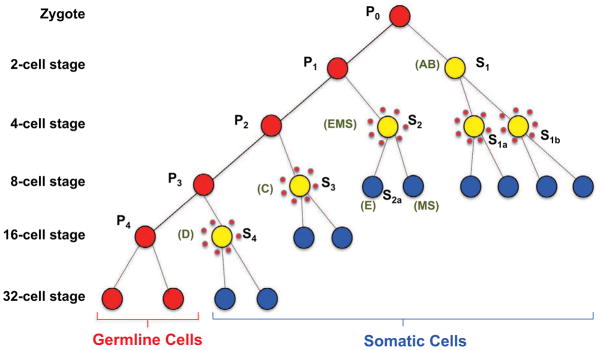
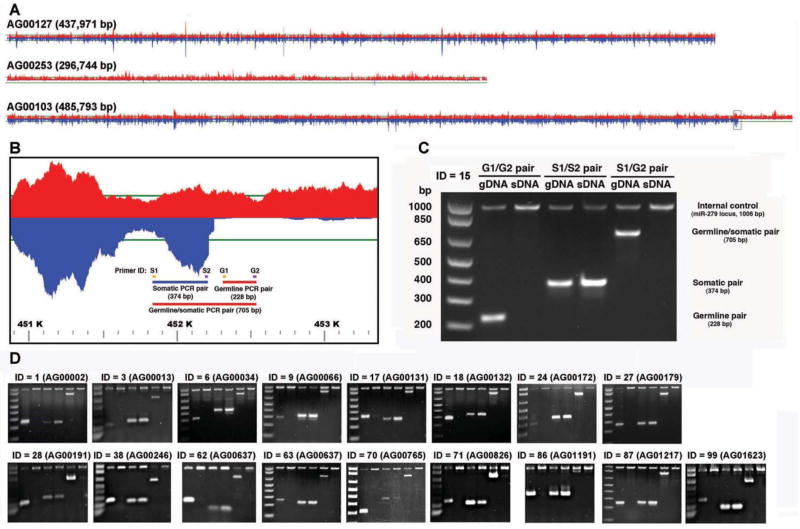
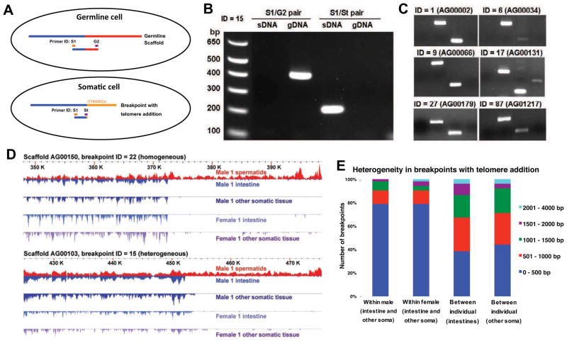
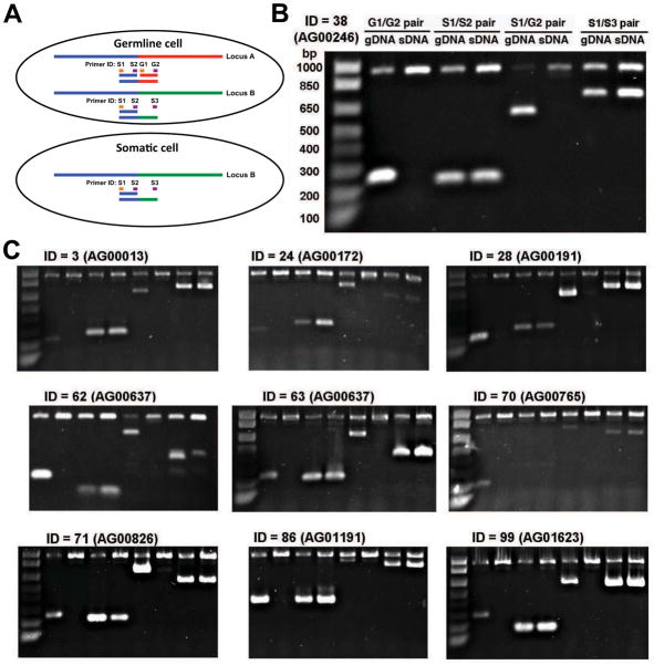
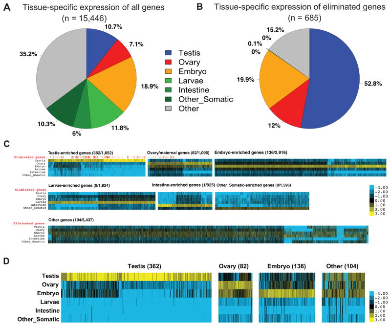
Chromatin diminution is the programmed elimination of specific DNA sequences during development. It occurs in diverse species, but the function(s) of diminution and the specificity of sequence loss remain largely unknown. Diminution in the nematode Ascaris suum occurs during early embryonic cleavages and leads to the loss of germline genome sequences and the formation of a distinct genome in somatic cells. We found that ∼43 Mb (∼13%) of genome sequence is eliminated in A. suum somatic cells, including ∼12.7 Mb of unique sequence. The eliminated sequences and location of the DNA breaks are the same in all somatic lineages from a single individual and between different individuals. At least 685 genes are eliminated. These genes are preferentially expressed in the germline and during early embryogenesis. We propose that diminution is a mechanism of germline gene regulation that specifically removes a large number of genes involved in gametogenesis and early embryogenesis.


Figures

Figure 1
Figure 1. A. suum early embryo development, cell lineage and chromatin diminution
Primordial germ cells (P) are in red, cells undergoing chromatin diminution are represented by yellow filled circles surrounded by dots, and blue cells (S) are precursor somatic cells and lineages. The primordial germ cells numbers correspond to their division state. P0 is the zygote whereas P1 through P4 represent the primordial germ cell derived from each subsequent cleavage of the germ cells as illustrated. S1–S4 cells are successive precursor somatic cell derived from each division of a germ cell (EMS = intestine, body wall muscle, and pharynx; E = intestine; MS = Body wall muscle, neurons, somatic gonad, coelomocytes, and pharynx; AB = Nervous system, hypodermis, and pharynx; C = Body wall muscle, hypodermis, and neurons; and D = Body wall muscle). Adapted from Theodor Boveri (Boveri, 1899, 1910) and Fritz Müller and Heinz Tobler (Goday and Pimpinelli, 1993; Muller et al., 1996; Muller and Tobler, 2000; Pimpinelli and Goday, 1989).
Figure 2
Figure 2. A. suum DNA elimination
A. Germline and somatic read coverage for regions of the A. suum genome illustrating the retention of segments in both the germline and somatic tissue (top), a region completely eliminated in the somatic tissue (middle), and a DNA breakpoint and region eliminated in the somatic tissues (bottom). Red designates germline reads and blue are somatic reads with the horizontal green line representing 50-fold coverage. B. Enlarged region of a scaffold (AG00103, Fig. 2A bottom) illustrating the PCR strategy used to verify DNA elimination predicted from the comparison of the germline and somatic genome sequences. C. PCR data confirm the elimination of DNA corresponding to scaffold AG00103 in A. suum somatic tissues. Note that the germline primer pair (G1/G2) produces a PCR product in the germline DNA (gDNA), but not somatic DNA (sDNA). The somatic primer pair (S1/S2) leads to PCR products in both the germline and somatic DNA, and the primer pair spanning the breakpoint (S1/G2) produces a PCR product only in the germline. gDNA = germline testis DNA and sDNA = somatic intestine DNA, isolated from the single male worm from which the genome sequences were derived. The 1,006 bp PCR product present in all lanes represents a control PCR corresponding to a single copy locus (miR-279) present in both the germline and somatic genomes. D. PCR data confirm the elimination of DNA in 17 additional independent loci in the A. suum somatic genome. The PCR strategy illustrated in B and C was applied to these loci. See also Figure S1 and Table S2.
Figure 3
Figure 3. A. suum chromosome breaks with telomere addition in somatic cells
A. The PCR strategy used to verify telomere sequence addition in the somatic cells. Primer St (Somatic telomere) is a hybrid primer consisting of 3′ nucleotides corresponding to the unique somatic sequence and 5′ nucleotides corresponding to telomeric sequence [(TTAGGC)n] (see Supplementary Experimental Procedures for primers sequences). B. PCR data confirm telomere addition in the A. suum somatic tissues at breakpoint 15. Primers are defined in Fig. 3A and genomic DNA sources defined as in Fig. 2C. C. PCR data confirm the telomere addition at 6 additional independent loci with chromosome breaks. D. Heterogeneity in breakpoints with telomere addition. Two breakpoints with telomere addition are illustrated. Note that the exact breakpoint for one of the loci in different somatic tissues varies (intestine and carcass), particularly between individuals. E. Overall heterogeneity in breakpoints with telomere addition. For these 52 breakpoints, we compared the genomes of pairs of somatic tissues (intestine and other somatic) from the same individual and between individuals and measured the difference in the position of the breakpoints identified. See also Figure S2 and Table S3.
Figure 4
Figure 4. Loss of one member of duplicated, rearranged loci
A. Loss of one of two similar germline loci in the somatic genome. Illustration shows two germline loci in a germline cell containing common sequences (> 97% identical) (blue line), divergent sequence (green or red lines), and the loss of one germline locus in somatic cells following chromatin diminution. Primers and PCR strategy used to verify loci in the germline and somatic genomes are shown. B. PCR data confirm locus A is present in germline cells but lost in somatic cells. Locus B is present in both germline and somatic cells. C. Additional PCR data for other loci demonstrating the loss of one member of duplicated, rearranged loci. See also Table S4.
Figure 5
Figure 5. Eliminated A. suum genes are primarily expressed in the germline
A. RNA expression of A. suum genes (n = 15,446) in different tissues. Gene expression enrichment was categorized by comparing RNA-seq data (Table S1) using reads per kilobase of template per million mapped reads (RPKM). B. The 685 eliminated A. suum genes are highly expressed in the germline and early embryogenesis. C. Expression Heatmap for all A. suum genes. Shown are expression heatmaps for different groups of genes illustrated in Fig. 5A. For each gene, the colors represent log2 values of fold changes to the average expression level (RPKM) for a gene in different stages. For each group of genes, the eliminated/total number of genes is indicated and a red vertical line above the heatmap marks the genes eliminated (see Fig. 5D). D. The expression profiles of eliminated A. suum genes. See Fig. 5C for legend. Note that for the 104 genes in the Other group, the majority of them are expressed in testis, ovary, and the early embryo. See also Figure S3 and Table S5–6.

Comment in

References

    1. Ashe A, Sapetschnig A, Weick EM, Mitchell J, Bagijn MP, Cording AC, Doebley AL, Goldstein LD, Lehrbach NJ, Le Pen J, et al. piRNAs Can Trigger a Multigenerational Epigenetic Memory in the Germline of C. elegans. Cell. 2012;150:88–99. - PMC - PubMed
    1. Bachmann-Waldmann C, Jentsch S, Tobler H, Muller F. Chromatin diminution leads to rapid evolutionary changes in the organization of the germ line genomes of the parasitic nematodes A. suum and P. univalens. Mol Biochem Parasitol. 2004;134:53–64. - PubMed
    1. Bagijn MP, Goldstein LD, Sapetschnig A, Weick EM, Bouasker S, Lehrbach NJ, Simard MJ, Miska EA. Function, targets, and evolution of Caenorhabditis elegans piRNAs. Science. 2012;337:574–578. - PMC - PubMed
    1. Boveri T. Ueber Differenzierung der Zellkerne wahrend der Furchung des Eies von Ascaris megalocephala. Anat Anz. 1887;2:688–693.
    1. Boveri T. Festschr fur C von Kupffer. Jena: Fisher; 1899. Die Entwicklung von Ascaris megalocephala mit besonderer Rucksicht auf die Kernverhaltnisse; pp. 383–430.

Publication types

Associated data