. 2013 Feb:240:75-87.
doi: 10.1016/j.expneurol.2012.10.023. Epub 2012 Nov 1.

Inhibition of the Jak-STAT pathway prevents CNTF-mediated survival of axotomized oxytocinergic magnocellular neurons in organotypic cultures of the rat supraoptic nucleus

Affiliations

Inhibition of the Jak-STAT pathway prevents CNTF-mediated survival of axotomized oxytocinergic magnocellular neurons in organotypic cultures of the rat supraoptic nucleus

Jason M Askvig et al. Exp Neurol. 2013 Feb.

Abstract

Previous studies have demonstrated that ciliary neurotrophic factor (CNTF) enhances survival and process outgrowth from magnocellular neurons in the paraventricular (PVN) and the supraoptic (SON) nuclei. However, the mechanisms by which CNTF facilitates these processes remain to be determined. Therefore, the aim of this study was to identify the immediate signal transduction events that occur within the rat SON following administration of exogenous rat recombinant CNTF (rrCNTF) and to determine the contribution of those intracellular signaling pathway(s) to neuronal survival and process outgrowth, respectively. Immunohistochemical and Western blot analyses demonstrated that axonal injury and acute unilateral pressure injection of 100 ng/μl of rrCNTF directly over the rat SON resulted in a rapid and transient increase in phosphorylated-STAT3 (pSTAT3) in astrocytes but not neurons in the SON in vivo. Utilizing rat hypothalamic organotypic explant cultures, we then demonstrated that administration of 25 ng/ml rrCNTF for 14days significantly increased the survival and process outgrowth of OT magnocellular neurons. In addition, pharmacological inhibition of the Jak-STAT pathway via AG490 and cucurbitacin I significantly reduced the survival of OT magnocellular neurons in the SON and PVN; however, the contribution of the Jak-STAT pathway to CNTF-mediated process outgrowth remains to be determined. Together, these data indicate that CNTF-induced survival of OT magnocellular neurons is mediated indirectly through astrocytes via the Jak-STAT signaling pathway.


Figures

Fig. 1
Fig. 1. Localization of tSTAT3 in the SON
Dual-label immunofluorescence experiments pairing anti-tSTAT3 (A, D, and G) with anti-OT (B), anti-VP (E), and anti-GFAP (H) revealed tSTAT3-immunoreactivity within OT (C) and VP (F) neurons and GFAP-immunoreactive astrocytes (I) of the SON (arrows). GFAP, glial fibrillary acidic protein; OC, optic chiasm; OT, oxytocin; tSTAT3, total STAT3; VP, vasopressin. Scale bar A-F = 75 μm; G-I = 150 μm.
Fig.2
Fig.2. pSTAT3 immunoreactivity in SON following infundibular nerve crush
(A) pSTAT3 immunoreactivity in the SON 3 hours post infundibular nerve crush demonstrated an increase in circular pSTAT3 immunoreactive profiles, confined to the SON-VGL and the SON (arrows). (B) Dual-label immunoperoxidase pairing anti-pSTAT3 (purple) with anti-GFAP (brown) confirmed the presence of pSTAT3 within GFAP-immunoreactive astrocytes in the parenchyma of the SON (arrows). Although obscured, pSTAT3-immunoreactivity can be discerned in the SON-VGL. Scale bar = 100 μm.
Fig. 3
Fig. 3. Localization of pSTAT3 in the SON following exogenous rrCNTF injection
At 1 hr post-pressure injection of 100 ng/μl rrCNTF, dual-label immunofluorescent experiments pairing anti-pSTAT3 (A, D) with anti-OT (B) and anti-GFAP (E) revealed complete co-localization of pSTAT3 with GFAP-immunoreactive astrocytes (F) in the ventral glial limitans of the SON (arrows) and the SON parenchyma (arrowheads), but not in OT (C, asterisks) or VP (not shown) magnocellular neurons. The inset in (F) clearly demonstrated pSTAT3-immunoreactivity within the GFAP-immunoreactive astrocytes in the SON. GFAP, glial fibrillary acidic protein; OC, optic chiasm; OT, oxytocin; pSTAT3, phosphorylated STAT3. Scale bar A-F = 25 μm; inset in F = 10 μm.
Fig. 4
Fig. 4. Exogenous rrCNTF resulted in the rapid and transient activation of STAT3 in the rat SON that is inhibited by AG490
(A) Western blot analysis of dissected SON samples demonstrated that acute pressure injection of 100 ng/μl rrCNTF induced a statistically significant increase in pSTAT3 levels in the SON at 1 hr post-CNTF-injection. At 3 hrs post-CNTF-injection pSTAT3 levels still remained significantly elevated compared to contralateral control and vehicle-infused control SON; however, the pSTAT3 levels at 3 hrs post-CNTF-injection were significantly decreased from the pSTAT3 levels at 1 hr post-CNTF-injection. Administration of AG490 (10 mg/kg IP) 1 hour prior to rrCNTF injection significantly decreased the pSTAT3 levels to levels that are not significantly different from vehicle-infused control SON but still significantly elevated from contralateral control SON. (B) Moreover, Western blot analysis demonstrated the level of pJak2 following AG490 inhibition is significantly decreased from all other groups (p<0.001), while the pJak2 levels in the control and rrCNTF injected SON do not differ significantly. Column bars and error bars represent the mean and SEM of 6 groups. Each group represents isolated SON pooled from three rats. In order to determine the amount of total protein that is phosphorylated, the ROD of the phosphorylated protein was normalized to the ROD of the total protein, as opposed to the ROD of ß-actin, to obtain a ratio used for statistical analysis. pJak2, phosphorylated Jak2; pSTAT3, phosphorylated STAT3; ROD, relative optical density; tJak2, total Jak2; tSTAT3, total STAT3.*p<0.05,**p<0.001, ***p<0.0001.
Fig. 5
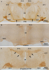
Fig. 5. Micrograph montages of magnocellular neurosecretory system nuclei in hypothalamic organotypic cultures immunohistochemically stained for OT magnocellular neurons
(A) Micrograph montage of magnocellular neurosecretory system nuclei of a representative explant slice fixed and immunohistochemically stained for OT magnocellular neurons at the time of sacrifice (post-natal day 6). Note the hypothalamo-neurohypophysial tract (A, arrows), which contains the magnocellular neuron axons that project to the NL. (B) Micrograph of OT magnocellular neurons of a representative explant slice that received control media for 14 days. Note the preservation of magnocellular neurosecretory system nuclei organization. (C) Montage of OT magnocellular neurons from a representative explant slice that received 25 ng/ml rrCNTF treatment for 14 days. Note the substantial increase in OT magnocellular neurons in the SON and PVN as well as the maintenance of process density following rrCNTF treatment (arrows) compared to control (B). Note that the representative images were obtained from approximately the same level of the magnocellular neurosecretory system, which is apparent when comparing the III ventricle (III) between the images. ACC, accessory nuclei; III, III ventricle; PVN, paraventricular nucleus; SON, supraoptic nucleus. Scale bar =500 μm.
Fig. 6
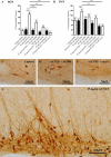
Fig. 6. The Jak-STAT pathway is necessary to mediate the CNTF-induced survival of OT neurons in the SON and PVN
Immunohistochemical neuronal cell counts demonstrated that exogenous rrCNTF promoted the survival of OT neurons while inhibition of the Jak-STAT pathway with AG490 or cucurbitacin I significantly reduced the number of surviving OT neurons in the SON (A) and PVN (B). Note the substantial increase in neuron numbers in the SON following rrCNTF administration (F) compared to the control media group (C), and the groups receiving the pharmacological inhibition of the Jak-STAT pathway (D, E). Column bars and error bars represent the mean and SEM of [n] groups. Cucu I, cucurbitacin I; PVN, paraventricular nucleus; SON, supraoptic nucleus. ***p<0.0001. Scale bar =100 μm.

References

    1. Albrecht PJ, Dahl JP, Stoltzfus OK, Levenson R, Levison SW. Ciliary neurotrophic factor activates spinal cord astrocytes, stimulating their production and release of fibroblast growth factor-2, to increase motor neuron survival. Exp Neurol. 2002;173:46–62. - PubMed
    1. Alonzi T, Middleton G, Wyatt S, Buchman V, Betz UA, Muller W, Musiani P, Poli V, Davies AM. Role of STAT3 and PI 3-kinase/Akt in mediating the survival actions of cytokines on sensory neurons. Mol Cell Neurosci. 2001;18:270–282. - PubMed
    1. Askvig JM, Leiphon LJ, Watt JA. Neuronal activity and axonal sprouting differentially regulate CNTF and CNTF receptor complex in the rat supraoptic nucleus. Exp Neurol. 2012;233:243–252. - PMC - PubMed
    1. Bechstein M, Haussler U, Neef M, Hofmann HD, Kirsch M, Haas CA. CNTF-mediated preactivation of astrocytes attenuates neuronal damage and epileptiform activity in experimental epilepsy. Exp Neurol. 2012;236:141–150. - PubMed
    1. Ben-Barak Y, Russell JT, Whitnall MH, Ozato K, Gainer H. Neurophysin in the hypothalamo-neurohypophysial system. I. Production and characterization of monoclonal antibodies. J Neurosci. 1985;5:81–97. - PMC - PubMed

Publication types

MeSH terms

LinkOut - more resources