. 2013 Jan 15;513(1):101-10.
doi: 10.1016/j.gene.2012.10.068. Epub 2012 Oct 31.

ADAR1 ablation decreases bone mass by impairing osteoblast function in mice

Affiliations

ADAR1 ablation decreases bone mass by impairing osteoblast function in mice

Shibing Yu et al. Gene. .

Abstract

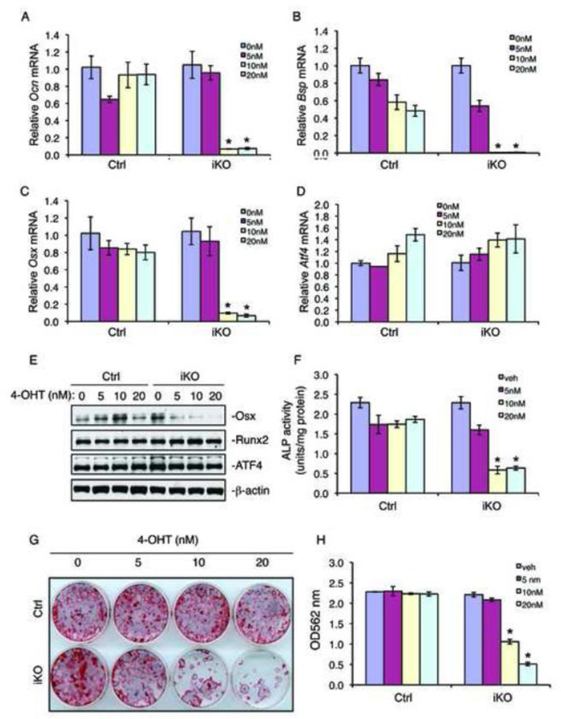
Bone mass is controlled through a delicate balance between osteoblast-mediated bone formation and osteoclast-mediated bone resorption. We show here that RNA editing enzyme adenosine deaminase acting on RNA 1 (ADAR1) is critical for proper control of bone mass. Postnatal conditional knockout of Adar1 (the gene encoding ADAR1) resulted in a severe osteopenic phenotype. Ablation of the Adar1 gene significantly suppressed osteoblast differentiation without affecting osteoclast differentiation in bone. In vitro deletion of the Adar1 gene decreased expression of osteoblast-specific osteocalcin and bone sialoprotein genes, alkaline phosphatase activity, and mineralization, suggesting a direct intrinsic role of ADAR1 in osteoblasts. ADAR1 regulates osteoblast differentiation by, at least in part, modulation of osterix expression, which is essential for bone formation. Further, ablation of the Adar1 gene decreased the proliferation and survival of bone marrow stromal cells and inhibited the differentiation of mesenchymal stem cells towards osteoblast lineage. Finally, shRNA knockdown of the Adar1 gene in MC-4 pre-osteoblasts reduced cyclin D1 and cyclin A1 expression and cell growth. Our results identify ADAR1 as a new key regulator of bone mass and suggest that ADAR1 functions in this process mainly through modulation of the intrinsic properties of osteoblasts (i.e., proliferation, survival and differentiation).


Conflict of interest statement

Disclosures: All the authors state that they have no conflicts of interest.

Figures

Figure 1
Figure 1. Postnatal conditional knockout of the Adar1 gene significantly decreases both trabecular and cortical bone in mice
(A) IHC staining. Five-µm tibial sections were immunohistochemically stained with an anti-ADAR1 antibody. Strong ADAR1 signal was detected in osteoblasts located on trabecular bone surfaces of Ctrl mice, which was lost in iKO osteoblasts. Original magnification: ×400. (BC) Representative images from 3D µCT reconstruction of femur trabecular (B) and cortical (C) bones. (DG) Quantitative analysis of trabecular number (Tb.N) (D), bone volume/tissue volume (BV/TV) (E), trabecular thickness (Tb.Th) (F), and cortical thickness (Cort.Th) (G) of femurs. N=4–6, *p <0.05 (versus Ctrl).
Figure 2
Figure 2. In vivo ablation of the Adar1 gene severely impairs osteoblast differentiation in bone and the ability of primary BMSCs from iKO mice to differentiate in vitro
(A) qPCR. Total RNA from Ctrl and iKO tibiae was used for qPCR using specific primers for Osx, Bsp, Osx, Runx2, and Atf4 mRNAs, which were normalized to Gapdh mRNA. N=4–6, *p <0.05 (versus Ctrl). (B) Western blot analysis. Protein extracts from Ctrl and iKO tibiae were used for Western blotting using specific antibodies against for Osx, Runx2, and ATF4 proteins. b-actin was used as a loading control. (C) IHC staining. Five-µm tibial sections were immunohistochemically stained with anti-Osx antibody or control IgG as indicated in the figure. Arrows indicate the nuclei of Osx-positive osteoblasts located on trabecular bone surfaces that were stained brown. Osx-negative cells were stained blue. Original magnification: ×100 (top), ×400 (bottom). (DF) BMSCs isolated from Ctrl and iKO mice were differentiated in vitro for 7d, followed by qPCR for Ocn, Osx, and Runx2 mRNAs (which was normalized to Gapdh mRNA) (D), or by Western blotting (E) for Osx and Runx2 (β-actin was used as a loading control), or by ALP activity assay (F). Bars represent means ± S.D. from three independent experiments. *p <0.05 (versus Ctrl). (G) Genomic DNAs were isolated from primary Ctrl and iKO BMSCs and used for PCR analysis using specific primers.
Figure 3
Figure 3. Ablation of the Adar1 gene does not alter osteoclast differentiation in bone
(AF) TRAP staining. Tibial sections of Ctrl and iKO mice were stained for TRAP activity. TRAP activity in metaphyseal regions of tibias is shown (A). Arrows indicate TRAP-positive osteoclasts on trabecular surfaces (B). Oc.S/BS and Oc.Nb/BPm in primary (C and D) and secondary (E and F) spongiosa of tibiae were measured. N=4–6. (G) IHC staining. Five-µm tibial sections of Ctrl and iKO mice were immunohistochemically stained with anti-Cat K antibody or control IgG as indicated in the figure. (H) qPCR. Tibiae from Ctrl and iKO mice were harvested for RNA isolation and qPCR analysis using specific primers for Cat K, Nfatc1, Rank, and Mmp9 mRNAs, which were normalized to Gapdh mRNA. N=4–6.
Figure 4
Figure 4. Ablation of the Adar1 gene dramatically decreases the formation of osteoblast progenitors in bone marrow cultures
(A and B) CFU-F assay. Bone marrow nucleated cells (1×10⁶ cells) from Ctrl and iKO mice were seeded in 35-mm culture dishes and cultured using the Mesencult Proliferation Kit (Mouse) for d10, followed by Giemsa staining. The numbers of CFU-Fs were counted under a microscope. (C and D) CFU-OB assay. Bone marrow nucleated cells (1×10⁶ cells) from Ctrl and iKO mice were seeded in 60-mm culture dishes in osteoblast differentiation medium (complete a-MEM containing 50 µg/ml L-ascorbic acid and 2.0 mM β-glycerophosphate). Media were changed every 2d. At d21, alizarin red staining was used to identify and enumerate the colonies containing mineralized bone matrix, which were designated as CFU-osteoblast (CFU-OB) colonies. Bars represent means ± S.D. from three independent experiments. *p<0.05 (versus Ctrl).
Figure 5
Figure 5. Ablation of the Adar1 gene decreases cell growth and increases cell apoptosis in primary BMSC cultures
(A) MTS assay. Primary Ctrl and iKO BMSCs were seeded at a density of 1×10⁴ cells/well in 96-well plates in proliferation medium. MTS assays were performed on days 0, 2, 4, 6, and 8 as indicated. *p<0.05 (vs. Ctrl). (BE) Ctrl and iKO BMSCs cultured in 8-well chambers (5×10⁵ cells/well) in proliferation medium for 72 h followed by BrdU staining (B and C) or TUNEL staining (D and E) as described in (Yu et al., 2009; Zhang et al., 2008). Arrows indicate BrdU-positive (proliferating) cells (B) or apoptotic cells (C). Magnification: 100X. Bars represent means ± S.D. from three independent experiments. *p <0.05 (vs. Ctrl).
Figure 6
Figure 6. shRNA knockdown of the Adar1 gene significantly decreases expression of cyclin D1 and cyclin A1 as well as cell growth in MC-4 pre-osteoblast cultures
(A–D) Adenoviral shRNA knockdown of ADAR1 in MC-4 cells. Cells were infected with equal amount of adenoviral vectors for ADAR1 shRNA or control shRNA. 48 h later, cells were harvested for Western blotting for cyclin A1, cyclin D1, p21 and p27. β-actin was used as a loading control (A), qPCR analysis for cyclin A1, cyclin D1, and cyclin D3 mRNAs, which were normalized to Gapdh mRNA (B), MTS assays (C), and direct cell count (D). Bars represent means ± S.D. from three independent experiments. *p <0.05 (vs. Control siRNA).
Figure 7
Figure 7. In vitro ablation of the Adar1 gene greatly impairs osteoblast differentiation in primary BMSC cultures
(AH) Osteoblast differentiation assays. Primary BMSCs isolated from Ctrl and iKO mice that were not previously treated with TM were treated with and without indicated concentrations of 4-hydroxytamoxifen (4OHT) for 24h in vitro and switched to osteoblast differentiation media for 7d (A–F), followed by RNA isolation for quantitative real-time PCR using specific primers for Ocn, Bsp, Osx, and Atf4 mRNAs, which were normalized to Gapdh mRNA (A–D), or for isolation of whole cell extracts for Western blot analysis using specific antibodies against for Osx, Runx2, and ATF4 proteins (β-actin was used as a loading control) (E), or for ALP assay (F). Primary BMSCs were treated with 4OHT as in (A–F) for 24h and switched to differentiation media for 14d, followed by alizarin red staining (G). Alizarin red was extracted with 10% cetylpyridinium chloride and OD562 was read (H). Bars represent means ± S.D. from three independent experiments. *p <0.05 (versus Ctrl).

References

    1. Agranat L, Raitskin O, Sperling J, Sperling R. The editing enzyme ADAR1 and the mRNA surveillance protein hUpf1 interact in the cell nucleus. Proc Natl Acad Sci U S A. 2008;105:5028–5033. - PMC - PubMed
    1. Banerjee C, McCabe LR, Choi JY, Hiebert SW, Stein JL, Stein GS, Lian JB. Runt homology domain proteins in osteoblast differentiation: AML3/CBFA1 is a major component of a bone-specific complex. J Cell Biochem. 1997;66:1–8. - PubMed
    1. Bass BL. RNA editing by adenosine deaminases that act on RNA. Annu Rev Biochem. 2002;71:817–846. - PMC - PubMed
    1. Burns CM, Chu H, Rueter SM, Hutchinson LK, Canton H, Sanders-Bush E, Emeson RB. Regulation of serotonin-2C receptor G-protein coupling by RNA editing. Nature. 1997;387:303–308. - PubMed
    1. Cao H, Yu S, Yao Z, Galson DL, Jiang Y, Zhang X, Fan J, Lu B, Guan Y, Luo M, Lai Y, Zhu Y, Kurihara N, Patrene K, Roodman GD, Xiao G. Activating transcription factor 4 regulates osteoclast differentiation in mice. J Clin Invest. 2010;120:2755–2766. - PMC - PubMed

Publication types

MeSH terms

LinkOut - more resources