. 2013 Jan;78(1):15-25.
doi: 10.1016/j.steroids.2012.10.002. Epub 2012 Nov 2.

DHEA metabolites activate estrogen receptors alpha and beta

Affiliations

DHEA metabolites activate estrogen receptors alpha and beta

Kristy K Michael Miller et al. Steroids. 2013 Jan.

Abstract

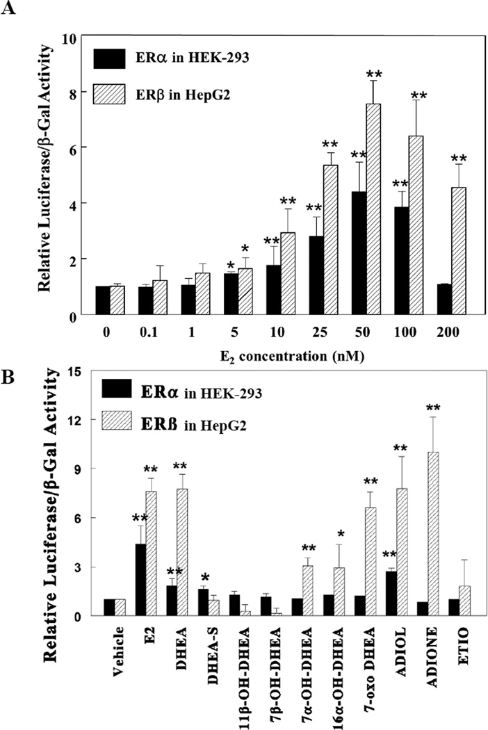
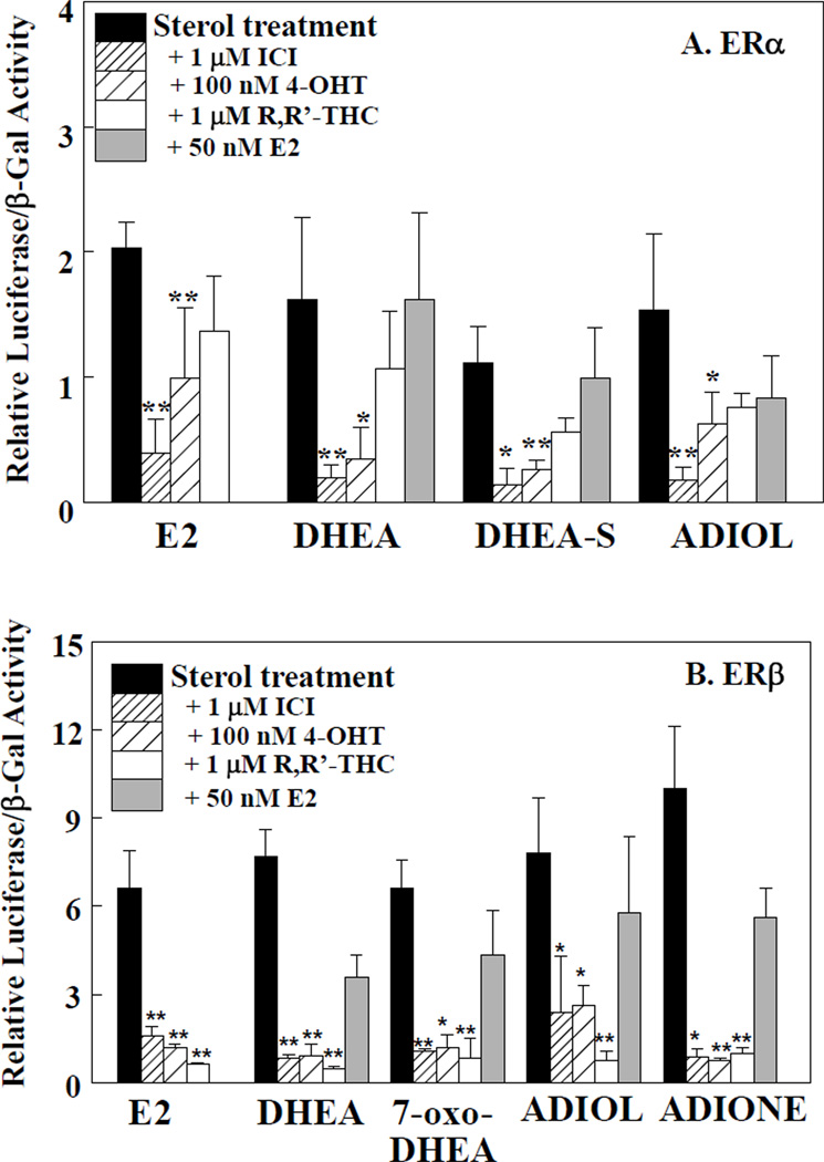
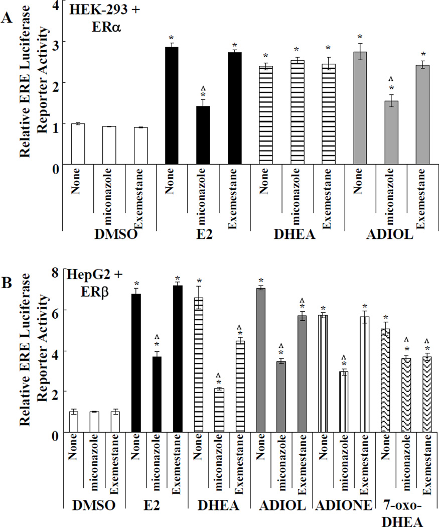
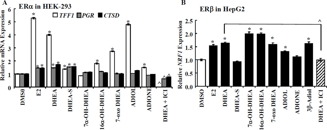
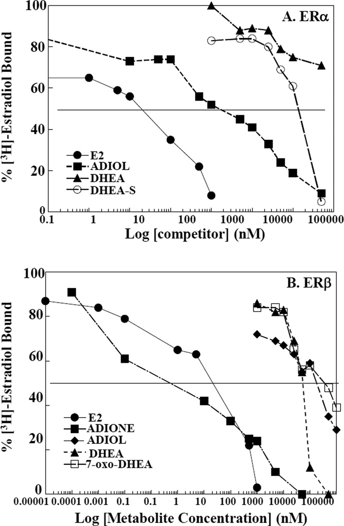
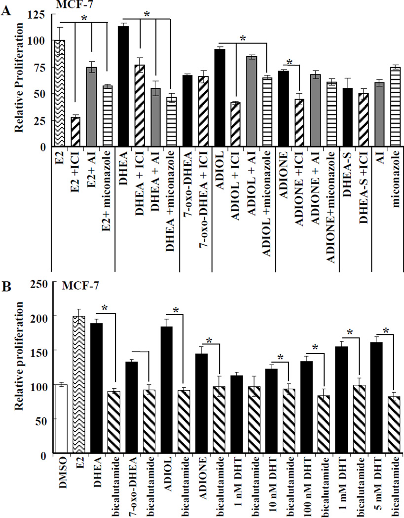
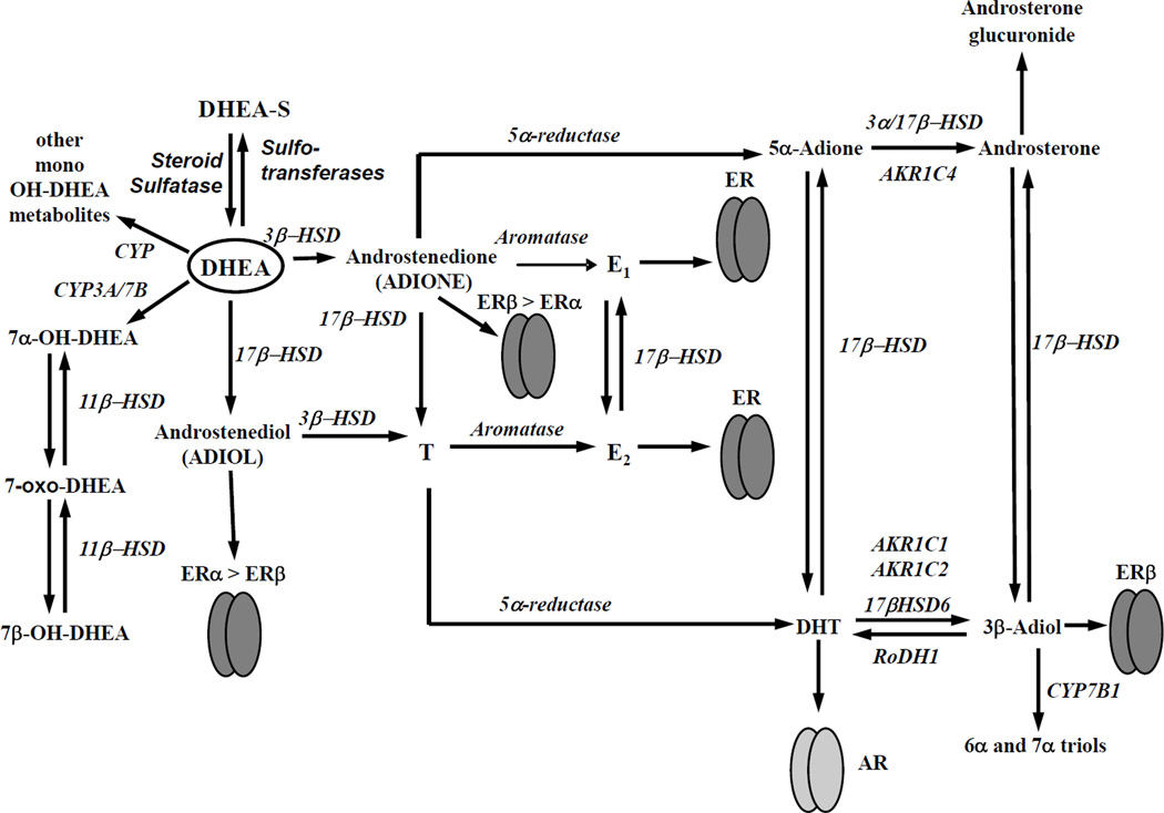
Dehydroepiandrosterone (DHEA) levels were reported to associate with increased breast cancer risk in postmenopausal women, but some carcinogen-induced rat mammary tumor studies question this claim. The purpose of this study was to determine how DHEA and its metabolites affect estrogen receptors α or β (ERα or ERβ)-regulated gene transcription and cell proliferation. In transiently transfected HEK-293 cells, androstenediol, DHEA, and DHEA-S activated ERα. In ERβ transfected HepG2 cells, androstenedione, DHEA, androstenediol, and 7-oxo DHEA stimulated reporter activity. ER antagonists ICI 182,780 (fulvestrant) and 4-hydroxytamoxifen, general P450 inhibitor miconazole, and aromatase inhibitor exemestane inhibited activation by DHEA or metabolites in transfected cells. ERβ-selective antagonist R,R-THC (R,R-cis-diethyl tetrahydrochrysene) inhibited DHEA and DHEA metabolite transcriptional activity in ERβ-transfected cells. Expression of endogenous estrogen-regulated genes: pS2, progesterone receptor, cathepsin D1, and nuclear respiratory factor-1 was increased by DHEA and its metabolites in an ER-subtype, gene, and cell-specific manner. DHEA metabolites, but not DHEA, competed with 17β-estradiol for ERα and ERβ binding and stimulated MCF-7 cell proliferation, demonstrating that DHEA metabolites interact directly with ERα and ERβin vitro, modulating estrogen target genes in vivo.


Figures

Figure 1
Figure 1. E2 activates ERE-luciferase activity in transiently transfected cells
HEK-293 cells were transfected with ERα and HepG2 were transfected with ERβ expression plasmids Both cell lines were cotransfected with ERELUC (estrogen-response element luciferase) and pCMV-β-gal reporters and were treated for 24 h with EtOH (vehicle) or A) the indicated concentrations of E2 or B) 10 nM E2, 5 µM DHEA or 5 µM of the indicated DHEA metabolites. The cells were harvested and the lysates were assayed for β-galactosidase and luciferase activities. Data represent the mean ± S.D. of three separate experiments. Statistical significance is indicated versus vehicle (EtOH control), * p<0.05, ** p<0.01.
Figure 2
Figure 2. Concentration-dependent activation of ERα by DHEA, DHEA-S, and ADIOL in ER transfected cells
HEK-293 and HepG2 cells were transfected with ERELUC reporter plasmid and expression vector for either human ERα (A) or ERβ (B), respectively. Cells were treated for 24 h with varying concentrations of E2, DHEA, DHEA-S, 7-oxo-DHEA, ADIONE and ADIOL. Cells were harvested and lysates were assayed for β-galactosidase and luciferase activities. Data represent the mean ± S.D. of three separate experiments. *, significantly different from vehicle-treated cells, p < 0.05, **,p<0.01. E2, ●; DHEA, ▲; DHEA-S, ○; ADIOL, ■; ADIONE, ♦; and 7-oxo-DHEA, □.
Figure 3
Figure 3. Inhibition of ERELUC reporter activity in the presence of cotransfected ERα in ER transfected cells
HEK-293 and HepG2 cells were transfected with an ERELUC reporter plasmid and an expression vector for either human ERα or ERβ, respectively. Cells were treated for 24 h with 5 µM DHEA metabolite in the absence or presence of either 1 µM 182,780 ICI, 100 nM 4-hydroxytamoxifen (4-OHT), 1 µM R,R-THC, or 50 nM 17β-estradiol (E2). Cells were then harvested and lysates assayed for β-galactosidase and luciferase activities. Data represent the mean ± S.D. of three wells. Experiments were repeated three times with similar results. A. ERα-mediated transactivation; B. ERβ-mediated transactivation. Statistical significance was determined using analysis of variance followed by Student's t tests. *, significantly different from treated cells, p < 0.05 *, or **p, 0.01.
Figure 4
Figure 4. Effect of non-selective P450 inhibitor miconazole or aromatase inhibitor (AI) exemestane on ER-mediated transcriptional activation by DHEA and metabolites
A) HEK-293 cells were transfected with ERα (A) and HepG2 cells were transfected with ERβ (B). All cells were co-transfected with ERELUC reporter plasmid and pRL-tk. After 24 h, cells were incubated for 24 h in phenol red-free MEM supplemented with 5% DCC-FBS plus DMSO, 10 nM E2, 5 µM DHEA, 5 µM ADIOL, 5 µM ADIONE, or 5 µM 7-oxo-DHEA, as indicated in each panel. Where indicated, cells were preincubated with 5 µM miconazole or 100 nM Exemestane (AI) for 6 h prior to addition of the hormone treatment. Values are the average of 3 separate determinations +/− SEM * Significantly different from DMSO (vehicle control), p < 0.05. ^ Significantly different from DHEA alone, p < 0.05.
Figure 5
Figure 5. DHEA and metabolites increase endogenous gene transcription
HEK-293 cells were transfected with ERα (A) and HepG2 were transfected with ERβ (B) expression plasmid for 24 h and then treated with the indicated hormones (10 nM E2 or 5 µM sterols) or 100 nM ICI 182,780 (ER antagonist) for 24 h. QPCR for A) TFF1 (pS2), PGR (PR), CTSD (cathepsin D) mRNA was normalized to 18S. B) NRF1 (NRF-1) was normalized to GAPDH. Values are the avg. ± SEM of triplicate determinations. * Significantly different from DMSO (vehicle control), p < 0.05. ^ Significantly different from DHEA alone, p < 0.05.
Figure 6
Figure 6. Competitive binding of DHEA metabolite to ERα and ERβ
An ligand binding competition assay using [3H] E2 was performed using baculovirus expressed ERα (A) or ERβ (B) with increasing concentrations of nonradiolabeled E2, DHEA or DHEA metabolites (DHEA-S, 7-oxo-DHEA, ADIONE and ADIOL). The values on the Y-axes are expressed as the percentage of [3H] E2 bound and each data point represents the mean of two independent binding assays. The competitor concentration causing 50% reduction in [3H] E2 binding (IC50) is found at the intersection of the binding curves with the 50% binding line (---------). E2, ●; DHEA, ▲; DHEA-S, ♦; ADIOL, ■; ADIONE, ♦; and 7-oxo-DHEA, □.
Figure 7
Figure 7. DHEA metabolites stimulate the proliferation of MCF-7 breast cancer cells
The effect of E2 (10 nM), and 5 µM of either DHEA, DHEA-S, 7-oxo-DHEA, ADIOL or ADIONE alone or in combination with 100 nM ICI 182,780 (fulvestrant, a pure ER antagonist, pretreatment for 6 h), 100 nM Exemestane (AI), or 5 µM miconzaole (A) or in the presence of 10 µM bicalutamide (Casodex, pretreatment for 4 h) on the proliferation of MCF-7 cells was measured by BrdU assay after 48 h treatment as described in Experimental. A) The absorbance values were converted to percent of E2 values for relative proliferation values. B) Values were normalized to DMSO vehicle control. The data are the mean ± S.E.M. of 3–4 independent experiments in which each treatment was performed in quadruplicate. *, Statistically different from that particular indicated ligand treatment alone; p < 0.05 (one-way ANOVA followed by Student, Newman, Keuls post-hoc testing).
Figure 8
Figure 8. Model of how DHEA metabolites activate ERα and ERβ
DHEA is converted to ADIOL, ADIONE, and mono-hydroxylated metabolites capable of activating the estrogen receptors. ADIOL and ADIONE are high affinity ligand activators of ER similar to the 3β-diol derived from DHT as demonstrated by Muthusamy et al [46]. Other DHEA metabolites, 7-hydroxy- and 7-keto-DHEA are low affinity ligand activators of ER, relative to ADIOL or ADIONE. Further information on the synthesis and metabolism of DHEA is reviewed in [, –92].

References

    1. Simpson ER, Davis SR. Minireview: aromatase and the regulation of estrogen biosynthesis-some new perspectives. Endocrinology. 2001;142:4589–4594. - PubMed
    1. Janni W, Hepp P. Adjuvant aromatase inhibitor therapy: Outcomes and safety. Cancer Treat Rev. 2010;36:249–261. - PubMed
    1. Kaaks R, Rinaldi S, Key TJ, Berrino F, Peeters PHM, Biessy C, et al. Postmenopausal serum androgens, oestrogens and breast cancer risk: the European prospective investigation into cancer and nutrition. Endocr Relat Cancer. 2005;12:1071–1082. - PubMed
    1. Danforth KN, Eliassen AH, Tworoger SS, Missmer SA, Barbieri RL, Rosner BA, et al. The association of plasma androgen levels with breast, ovarian and endometrial cancer risk factors among postmenopausal women. Int J Cancer. 2010;126:199–207. - PMC - PubMed
    1. Endogenous H Breast Cancer Collaborative G. Circulating sex hormones and breast cancer risk factors in postmenopausal women: reanalysis of 13 studies. Br J Cancer. 2011 - PMC - PubMed

Publication types

MeSH terms