. 2012 Dec 7;531(2):125-30.
doi: 10.1016/j.neulet.2012.10.036. Epub 2012 Nov 2.

Rit GTPase regulates a p38 MAPK-dependent neuronal survival pathway

Affiliations

Rit GTPase regulates a p38 MAPK-dependent neuronal survival pathway

Weikang Cai et al. Neurosci Lett. .

Abstract

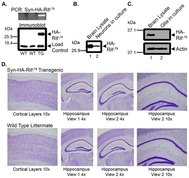
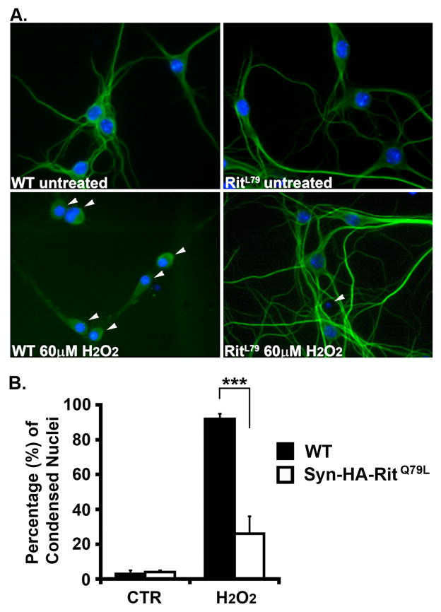
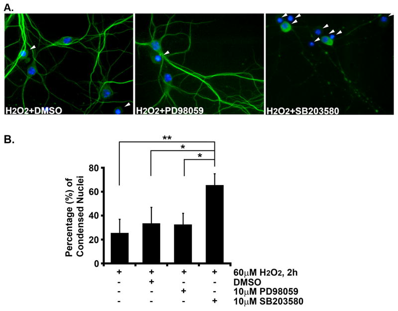
Rit, along with Rin and Drosophila Ric, comprises the Rit subfamily of Ras-related small GTPases. Although the cellular functions of many Ras family GTPases are well established, the physiological significance of Rit remains poorly understood. Loss of Rit sensitizes multiple mammalian cell lines and mouse embryonic fibroblasts (MEFs) derived from Rit(-/-) mice to oxidative stress-mediated apoptosis. However, whether Rit-mediated pro-survival signaling extends to other cell types, particularly neurons, is presently unknown. Here, to examine these issues we generated a transgenic mouse overexpressing constitutively active Rit (Rit(Q79L)) exclusively in neurons, under control of the Synapsin I promoter. Active Rit-expressing hippocampal neurons display a dramatic increase in oxidative stress resistance. Moreover, pharmacological inhibitor studies demonstrate that p38 MAPK, rather than a MEK/ERK signaling cascade, is required for Rit-mediated protection. Together, the present studies identify a critical role for the Rit-p38 MAPK signaling cascade in promoting hippocampal neuron survival following oxidative stress.


Figures

Fig. 1
Fig. 1. HA-RitQ79L protein under the control of the Synapsin I promoter is expressed exclusively in neurons and does not alter brain morphology
(A) Upper panel, PCR analysis of wild-type (WT) or SynCARit (TG) animals screened with primers for the human Rit gene. Lower panel, anti-HA immunoblotting was used to detect HA-RitQ79L expression from brain lysate (100 μg) generated from the same animals as in the Upper panel. The non-specific band serves as a loading control. (B) Neurons were harvested and subjected to anti-HA immunoblotting (100 μg). Whole brain lysate (100 μg) served as positive control. (C) Mixed glial cell cultures (100 μg) were subjected to anti-HA immunoblotting. (D) Coronal sections from adult SynCARit or WT littermates were Nissl stained and images captured with an AX80 light microscope.
Fig. 2
Fig. 2. SynRit neurons are protected from ROS-induced death
(A) Hippocampal neurons (DIV8) isolated from WT or SynCARit P1 mouse pups were left untreated or treated with H2O2 (60 μM) for 2h and coimmunostained with MAP2 antibody (green) and Hoechst stain (blue). Arrowheads indicate neurons with condensed nuclei. (B) The percentage of MAP2+ cells with condensed nuclei are presented as mean ± SEM. Experiments were performed in triplicate. *** p<0.0001 using Fisher’s PLSD post-hoc test.
Fig. 3
Fig. 3. Inhibition of p38 MAPK but not ERK blocks RitQ79L-mediated protection from reactive oxygen species-induced death
(A) Hippocampal neurons (DIV8) were pre-treated for 30 min with DMSO, PD98059, or SB203580, and subjected to H2O2 exposure (60 μM) for 2 h. Cells were fixed, stained (neurons, MAP2, green; nuclei, Hoechst, blue) and scored for condensed nuclei (arrowhead). Representative images are shown. (B) The percentage of MAP2+ cells with condensed nuclei are presented as mean ± SEM. Experiments were performed in triplicate. * p<0.02, ** p<0.006 using Fisher’s PLSD post-hoc test.

References

    1. Andres DA, Shi GX, Bruun D, Barnhart C, Lein PJ. Rit signaling contributes to interferon-gamma-induced dendritic retraction via p38 mitogen-activated protein kinase activation. Journal of neurochemistry. 2008;107:1436–1447. - PMC - PubMed
    1. Arthur JS, Fong AL, Dwyer JM, Davare M, Reese E, Obrietan K, Impey S. Mitogen- and stress-activated protein kinase 1 mediates cAMP response element-binding protein phosphorylation and activation by neurotrophins. The Journal of neuroscience. 2004;24:4324–4332. - PMC - PubMed
    1. Cai W, Carlson SW, Brelsfoard JM, Mannon CE, Moncman CL, Saatman KE, Andres DA. Rit GTPase Signaling Promotes Immature Hippocampal Neuronal Survival. Journal of Neuroscience. 2012;32:9887–9897. - PMC - PubMed
    1. Cai W, Rudolph JL, Harrison SM, Jin L, Frantz AL, Harrison DA, Andres DA. An evolutionarily conserved Rit GTPase-p38 MAPK signaling pathway mediates oxidative stress resistance. Molecular biology of the cell. 2011;22:3231–3241. - PMC - PubMed
    1. Campbell SL, Khosravi-Far R, Rossman KL, Clark GJ, Der CJ. Increasing complexity of Ras signaling. Oncogene. 1998;17:1395–1413. - PubMed

Publication types

Substances

LinkOut - more resources