. 2013 Jan:54:1-16.
doi: 10.1016/j.freeradbiomed.2012.10.553. Epub 2012 Nov 1.

IL-4 and IL-13 employ discrete signaling pathways for target gene expression in alternatively activated monocytes/macrophages

Affiliations

IL-4 and IL-13 employ discrete signaling pathways for target gene expression in alternatively activated monocytes/macrophages

Ashish Bhattacharjee et al. Free Radic Biol Med. 2013 Jan.

Abstract

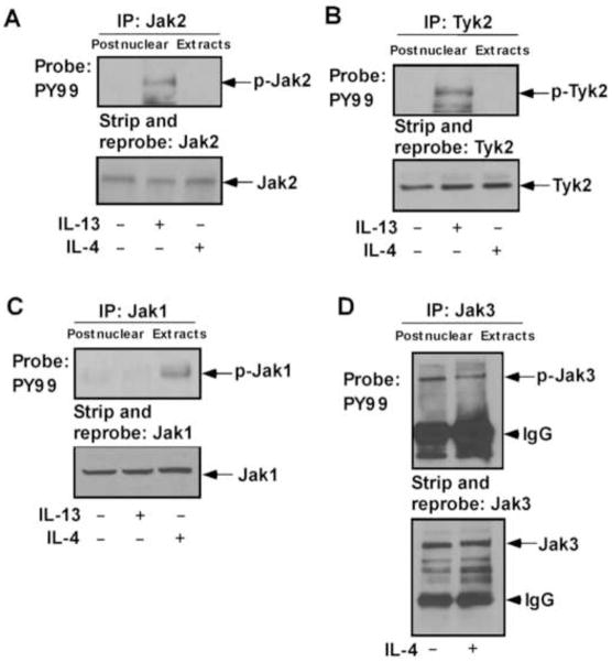
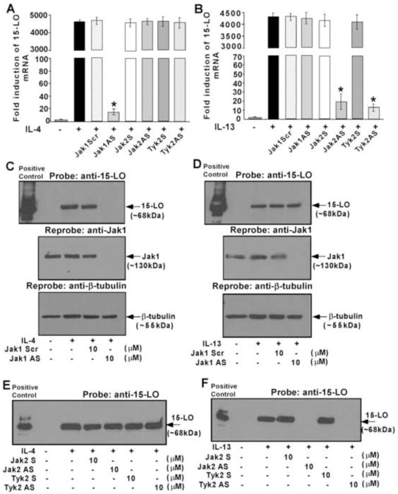
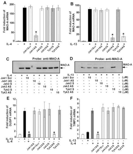
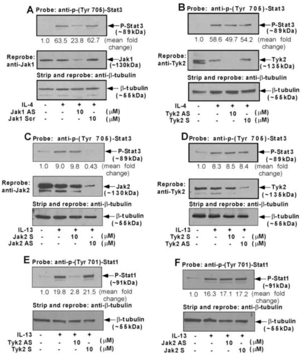
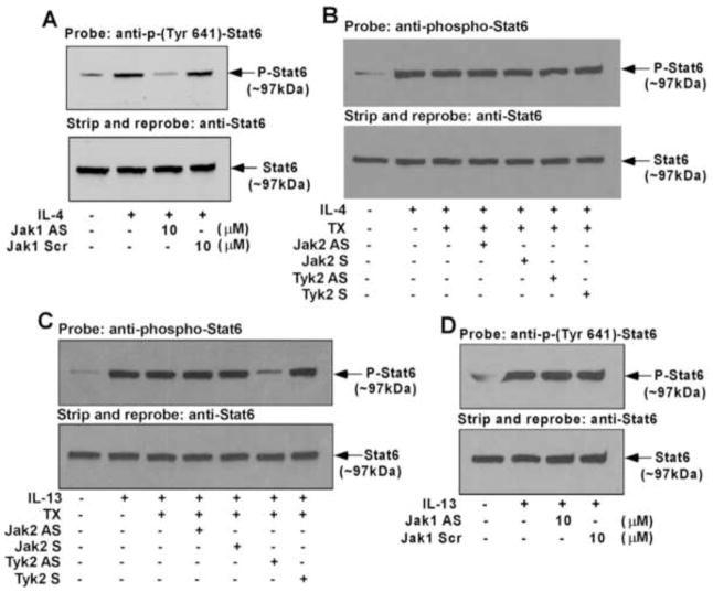
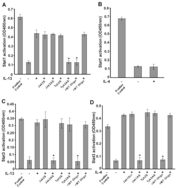
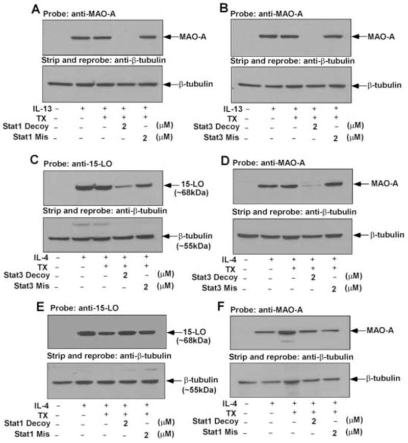
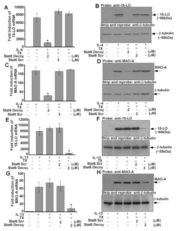
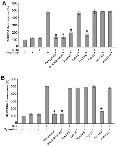
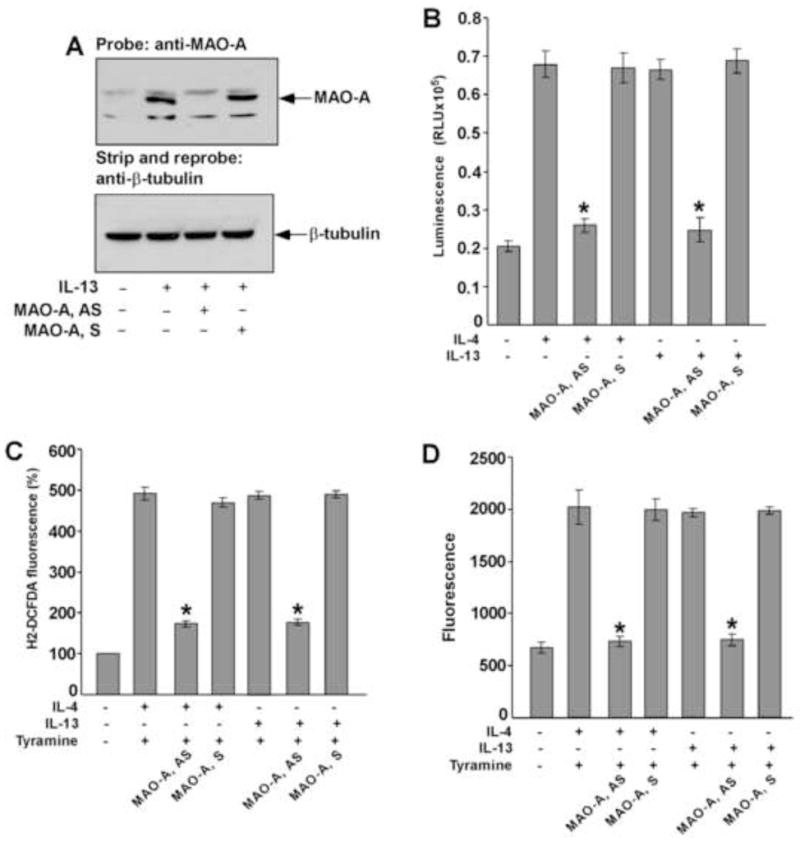
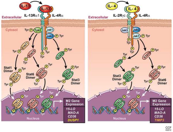
Monocytes/macrophages are innate immune cells that play a crucial role in the resolution of inflammation. In the presence of the Th2 cytokines interleukin-4 (IL-4) and interleukin-13 (IL-13), they display an anti-inflammatory profile and this activation pathway is known as alternative activation. In this study we compare and differentiate pathways mediated by IL-4 and IL-13 activation of human monocytes/macrophages. Here we report differential regulation of IL-4 and IL-13 signaling in monocytes/macrophages starting from IL-4/IL-13 cytokine receptors to Jak/Stat-mediated signaling pathways that ultimately control expression of several inflammatory genes. Our data demonstrate that although the receptor-associated tyrosine kinases Jak2 and Tyk2 are activated after the recruitment of IL-13 to its receptor (containing IL-4Rα and IL-13Rα1), IL-4 stimulates Jak1 activation. We further show that Jak2 is upstream of Stat3 activation and Tyk2 controls Stat1 and Stat6 activation in response to IL-13 stimulation. In contrast, Jak1 regulates Stat3 and Stat6 activation in IL-4-induced monocytes. Our results further reveal that although IL-13 utilizes both IL-4Rα/Jak2/Stat3 and IL-13Rα1/Tyk2/Stat1/Stat6 signaling pathways, IL-4 can use only the IL-4Rα/Jak1/Stat3/Stat6 cascade to regulate the expression of some critical inflammatory genes, including 15-lipoxygenase, monoamine oxidase A (MAO-A), and the scavenger receptor CD36. Moreover, we demonstrate here that IL-13 and IL-4 can uniquely affect the expression of particular genes such as dual-specificity phosphatase 1 and tissue inhibitor of metalloprotease-3 and do so through different Jaks. As evidence of differential regulation of gene function by IL-4 and IL-13, we further report that MAO-A-mediated reactive oxygen species generation is influenced by different Jaks. Collectively, these results have major implications for understanding the mechanism and function of alternatively activated monocytes/macrophages by IL-4 and IL-13 and add novel insights into the pathogenesis and potential treatment of various inflammatory diseases.


Conflict of interest statement

The authors have no financial conflict of interest.

Figures

Figure 1
Figure 1. IL-4 and IL-13 activates different Jak kinases in monocytes
Human blood monocytes (10×106/group) were directly stimulated with IL-13 or IL-4 for 10 min or left untreated as indicated. Cells were lysed and the postnuclear extracts were immunoprecipitated (IP) with different Jak/Tyk antibodies as mentioned in panels A–D. The immune complexes were analyzed by immunoblotting with antibody to phosphotyrosine, PY99 and presented in the upper panels of A–D. The bottom panels represent stripping and reprobing the blot with the same individual antibody used for immunoprecipitation, to assess equal protein loading. Arrows indicate the positions of respective Jak kinases as mentioned, based on the migration of molecular weight markers that were run in adjacent lanes. The arrowhead marks the migration of the heavy chain of IgG. Data are from a representative experiment of three independent experiments that were performed.
Figure 2
Figure 2. Induction of 15-LO expression by IL-4 and IL-13 requires different Jak kinases in monocytes
Monocytes (5×10⁶/group) were pre-treated directly with antisense ODN to Jak1 or a scrambled ODN control (10 μM) (A, B, C, D) or Jak2 or Tyk2 antisense or sense ODNs (10 μM) (A, B, E, F) for 48 h, with one re-feed at 24 h, prior to the incubation with IL-4 (A, C, E) or IL-13 (B, D, F) for another 24h. In panels A–B, total cellular RNA extracts were prepared and subjected to real-time quantitative PCR analysis. After normalization with GAPDH amplification, the fold induction of 15-LO mRNA expression for different groups was plotted. Data are the means ± S.D. (n=3). Significant differences were determined by comparing the antisense (AS), scrambled (Scr) and sense (S) ODN treated groups to the IL-4 or IL-13-treated control (*p<0.002). In panels C–F, cells were lysed and 50μg of postnuclear extracts were separated by SDS-PAGE. The proteins were resolved, transferred to a PVDF membrane and immunoblotted with an antibody against 15-LO (upper panels of C and D, E, F). The same blots were then reprobed with Jak1 antibody to examine the effect of antisense ODN on Jak1 protein expression (middle panels of C and D). To confirm equal loading the blots were then stripped and reprobed with (β-tubulin antibody (lower panels of C and D). Arrows indicate the predicted migration of 15-LO, Jak1 and β-tubulin as determined by the migration of molecular weight markers in adjacent lanes. Recombinant 15-LO was used as a positive control. The data shown represent one of three separate experiments giving similar results.
Figure 3
Figure 3. IL-4 and IL-13-stimulated MAO-A expression as well as IL-13 and IL-4 specific induction of DUSP1 and TIMP3 is controlled by different Jak kinases in human monocytes/macrophages
Monocytes (5×10⁶/group) were pre-treated directly with antisense ODN to Jak1 or a scrambled ODN control (10 μM) (A–F) or Jak2 or Tyk2 antisense or sense ODNs (10 μM) (A–F) for 48 h, with one re-feed at 24 h, prior to the incubation with IL-4 (A, C, E, F) or IL-13 (B, D, E, F) for another 24h (A–D) or 48h (E, F). In panels A–B, total cellular RNA extracts were prepared and subjected to real-time quantitative PCR analysis. After normalization with GAPDH amplification, the fold induction of MAO-A mRNA expression for different groups was plotted. Data are the means ± S.D. (n=3). Significant differences were determined by comparing the antisense (AS), scrambled (Scr) and sense (S) ODN treated groups to the IL-4 or IL-13-treated control (*p<0.002) In panels C–D, cells were lysed and 50μg of postnuclear extracts were separated by SDS-PAGE and immunoblotted with an antibody against MAO-A. The data shown are representative of three separate experiments giving similar results. For panels E and F, real-time PCR analyses was performed and fold induction of DUSP1 (E) and TIMP3 (F) mRNA expression for different groups was plotted after normalization with GAPDH. Data are the mean ± SD; (n=3). *p<0.003 compared to the IL-13 (E) or IL-4 (F)-treated controls.
Figure 4
Figure 4. Jak2 and Tyk2 are the upstream regulators of Stat3 and Stat1 activation in IL-13-stimulated monocytes whereas Jak1 controls Stat3 activation in IL-4-treated monocytes
Monocytes (5×10⁶/group) were either pre-treated with antisense ODN to Jak1 or scrambled ODN control (A), Tyk2 antisense or sense ODNs (B, D, E) or with Jak2 antisense or sense ODNs (C, F) for 48 h, with one re-feed at 24 h, prior to the addition of IL-4 (A, B) and IL-13 (C–F) for 15 min. The cells were lysed and whole cell extracts were resolved by SDS-PAGE. Stat3 and Stat1 tyrosine phosphorylation was detected by immunoblotting using p-Tyr705 Stat3 (upper panels of A–D) and p-Tyr701 Stat1 (upper panels of E and F) antibodies. The same blots were then reprobed with Jak1 (middle panel of A), Tyk2 (middle panels of B and D) and Jak2 (middle panel of C) antibodies to examine the effect of antisense ODN on Jak1, Tyk2 and Jak2 protein expression levels. To assess equal loading the blots were then stripped and reprobed with (β-tubulin antibody (lower panels of A–F). The data shown are representative of three independent experiments giving similar results. Blots from three different experiments were quantified and normalized to the respective total protein loads (β-tubulin). Mean fold change data are provided for all panels after considering all untreated groups (no IL-4 and IL-13) as 1.0.
Figure 5
Figure 5. IL-4-stimulated Stat6 activation is mediated by Jak1 whereas Tyk2 is required for Stat6 activation in IL-13-treated monocytes
Monocytes (5×10⁶/group) were pre-treated with scrambled/sense control ODNs or antisense ODNs to Jak1 (A, D) or Jak2 or Tyk2 (B, C) according to protocols described under “Materials and Methods” prior to the addition of IL-4 (A, B) or IL-13 (C, D) for 30 min. The cells were lysed and immunoblotted with anti-phospho-Stat6 antibody (upper panels of A–D). The same blots were stripped and reprobed with an antibody recognizing total Stat6 to confirm equal loading (lower panels of A–D). Data in panels A–D are from a representative of three repeat experiments showing similar results.
Figure 6
Figure 6. IL-13-induced Stat1 DNA binding activity requires Tyk2 whereas Jak1 and Jak2 are involved in regulating IL-4 and IL-13-induced Stat3 DNA binding activity in human monocytes
Human monocytes (5×10⁶/group) were either directly exposed to IL-13 (A, C) or IL-4 (B, D) for 1h or pretreated with control ODN or antisense ODN to Jak2 or Tyk2 (A, C) or Jak1 or Tyk2 (D) (10 μM) for 48 h, with one re-feed at 24 h followed by IL-13 or IL-4 stimulation for 1h. Nuclear extracts (5μg) were run in duplicate to perform an immunodetection of activated Stat1 (A, B) and Stat3 (C, D) using a TransAM Stat family kit. Nuclear extracts from IFNγ-stimulated COS-7 cells and IL-6-stimulated HepG2 cells were used as positive controls for activated Stat1 (A, B) and activated Stat3 (C, D) respectively. The wild-type (WT) and mutated (MT) consensus oligonucleotides were used to monitor the specificity of the assay. Values are mean ± SEM of three separate experiments giving similar results. Significant differences were determined by comparing each group to the IL-13 or IL-4-treated monocytes as the control (*p<0.004).
Figure 7
Figure 7. IL-13 stimulation requires both Stat1 and Stat3 whereas only Stat3 is required by IL-4 for the expression of 15-LO and MAO-A in alternatively activated monocytes
Monocytes (5×10⁶/group) were transfected with or without decoy or mismatched decoy ODNs to Stat1 (A, E, F) or Stat3 (B, C, D) according to protocols described under “Materials and Methods” prior to the addition of IL-13 (A, B) or IL-4 (C–F) for 48 h. Monocyte lysates were resolved by SDS-PAGE and immunoblotted with MAO-A (upper panels of A, B, D, F) or 15-LO (upper panels of C and E) specific antibodies. The same blots were then stripped and reprobed with (β-tubulin antibody (lower panels of A–F) to assess equal loading. Data are from a representative of three repeat experiments showing similar results.
Figure 8
Figure 8. Stat6 regulates 15-LO and MAO-A gene expression in IL-4 and IL-13-activated monocytes
Human blood monocytes (5×10⁶/group) were transfected with or without decoy or scrambled ODN to Stat6 according to protocols described under “Materials and Methods” prior to the addition of IL-4 (A–D) or IL-13 (E–H) for 24 h. 15-LO (A, E) and MAO-A (C, G) mRNA expression was detected by real time quantitative PCR analysis. Data are the mean ± SD; (n=3). Significant differences were determined by comparing the decoy or scrambled decoy ODN (to Stat6) treated groups to the transfection control (IL-4 or IL-13-treated) (*p<0.006). In B, D, F and H, monocyte lysates were separated by SDS-PAGE and immunoblotted with 15-LO (upper panels of B and F) and MAO-A (upper panels of D and H) antibodies. The same blots were then stripped and reprobed with (β-tubulin antibody (lower panels of B, D, F and H) to assess equal loading. Data in panels A–H are from a representative of three independent experiments showing similar results.
Figure 9
Figure 9. Tyramine-induced reactive oxygen species (ROS) generation is controlled by different Jak kinases in IL-13 versus IL-4 activated human monocytes/macrophages
Monocytes (5×10⁶/group) were pre-treated with control ODN or antisense ODNs to Jak1 or Jak2 or Tyk2 (A, B) according to protocols described under “Materials and Methods” or with the MAO inhibitors, pargyline (1 μM) (A, B) and Moclobemide (1 μM) (A, B) for 30 min prior to stimulation with IL-13 (2nM) (A) or IL-4 (2nM) (B) for 24h. Monocytes/macrophages were stimulated by tyramine (5 μM) for 30 min and then incubated with the fluorescent probe H2DCFDA (5 μM) for another 30 min before the fluorescence was measured (A, B). Data in panels A and B are expressed as percentage of unstimulated controls and represented as mean ± SD; (n=3, *p<0.004). Data are from a representative of three independent experiments.
Figure 10
Figure 10. MAO-A regulates tyramine-induced ROS generation in IL-13 and IL-4-activated monocytes/macrophages
Monocytes (5×10⁶/group) were pre-treated with MAO-A sense (S) or antisense (AS) ODN (A–D) according to protocols described under “Materials and Methods” and incubated with IL-13 (A–D) and IL-4 (B–D) (2nM) for 46h. In panel A, whole cell extracts (50 μg/lane) were resolved by SDS-PAGE. MAO-A protein expression was detected on Western blots with a MAO-A specific antibody (upper panel of A). The blot was stripped and reprobed with an antibody against (β-tubulin (lower panel of A) to assess equal loading. In panel B, native MAO-A enzyme activity was detected. In a total volume of 50 μl, 20 μl whole cell extract was incubated with substrate (final conc. 40 μM) in presence of MAO reaction buffer [100mM HEPES (pH 7.5); 5% glycerol]. After incubation at room temperature for 3h, 50μl luciferin detection reagent was added and the luminescent signal was measured after 20 min. Data represented as mean ± SD; (n=3, *p<0.002). In panel C, monocytes/macrophages were stimulated by tyramine (5 μM) for 30 min and then incubated with the fluorescent probe H2DCFDA (5 μM) for another 30 min before the fluorescence was measured. Data in panel C is expressed as percentage of unstimulated controls and represented as mean ± SD; (n=3, *p<0.004). In panel D, monocyte lysates (50μg/well) were incubated with Amplex Red reagent/HRP/p-Tyramine working solution for 60 min before the fluorescence was measured. Data represented as mean ± SD; (n=3, *p<0.005). Data in all panels are from a representative of three independent experiments.
Figure 11
Figure 11. Proposed model showing the differential regulation of gene expression in alternatively activated monocytes/macrophages by IL-13 and IL-4
Schematic representations indicate that both IL-13 and IL-4 share and signal through a common receptor component IL-4Rα. For IL-13 the other receptor component is IL-13Rα1 whereas the IL-4 receptor contains IL-2Rγc. These receptor components are phosphorylated upon exposure of monocytes to IL-13 or IL-4. Different Jak kinases are associated with these receptor components and activated (tyrosine phosphorylated) in response to IL-13 or IL-4 stimulation as presented. Consequently Stat1 (for IL-13 only), Stat3 and Stat6 (for both IL-13 and IL-4) are activated, form dimmers and, translocate into the nucleus, bind DNA and facilitate the induction of 15-LO, MAO-A and CD36 gene expression. IL-13 and IL-4 differentially regulate the gene expression of DUSP1 and TIMP3 through distinct Jak/Stat pathways. Different color code for DUSP1 and TIMP3 is used to demonstrate the unique regulation of these genes by IL-13 and IL-4 respectively.

References

    1. Iademarco MF, Barks JL, Dean DC. Regulation of vascular cell adhesion molecule-1 expression by IL-4 and TNF-alpha in cultured endothelial cells. J Clin Invest. 1995;95:264–271. - PMC - PubMed
    1. Miossec P, van den Berg W. Th1/Th2 cytokine balance in arthritis. Arthritis Rheum. 1997;40:2105–2115. - PubMed
    1. Gordon S. Alternative activation of macrophages. Nat Rev Immunol. 2003;3:23–35. - PubMed
    1. Folcik VA, Aamir R, Cathcart MK. Cytokine modulation of LDL oxidation by activated human monocytes. Arterioscler Thromb Vasc Biol. 1997;17:1954–1961. - PubMed
    1. Allen JB, Wong HL, Costa GL, Bienkowski MJ, Wahl SM. Suppression of monocyte function and differential regulation of IL-1 and IL-1ra by IL-4 contribute to resolution of experimental arthritis. J Immunol. 1993;151:4344–4351. - PubMed

Publication types

MeSH terms

LinkOut - more resources