. 2013 Jan 4;288(1):99-109.
doi: 10.1074/jbc.M112.416354. Epub 2012 Nov 2.

The human W42R γD-crystallin mutant structure provides a link between congenital and age-related cataracts

Affiliations

The human W42R γD-crystallin mutant structure provides a link between congenital and age-related cataracts

Fangling Ji et al. J Biol Chem. .

Abstract

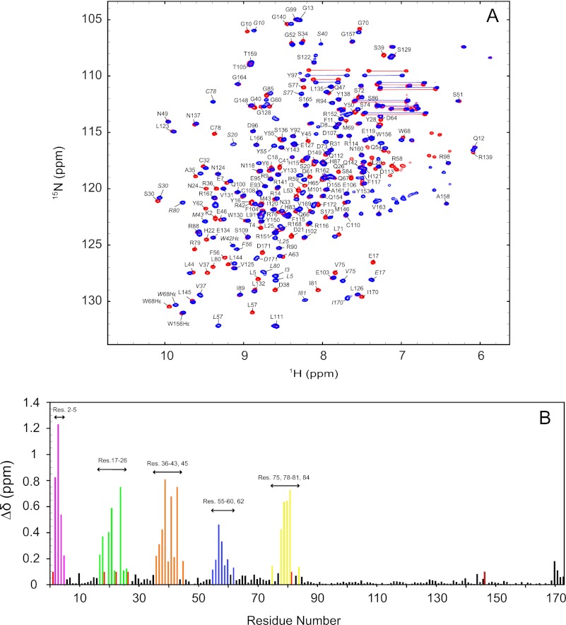
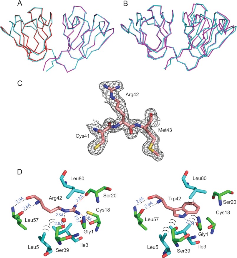
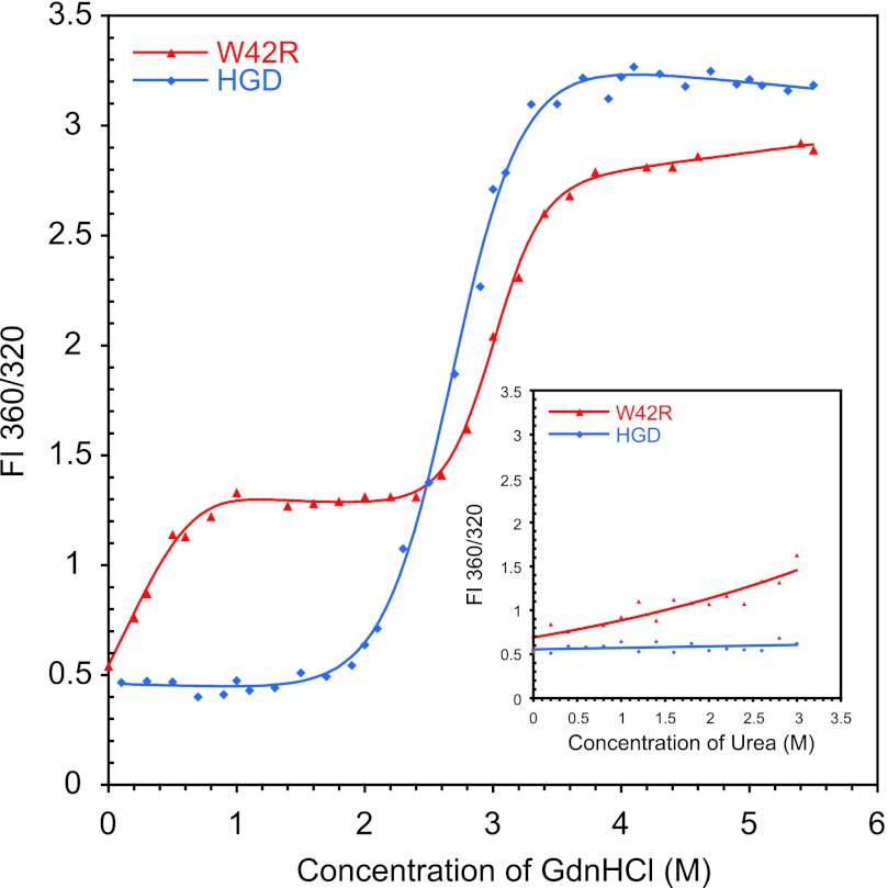
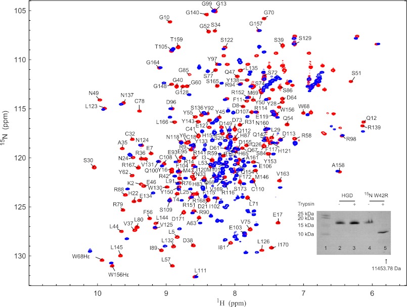
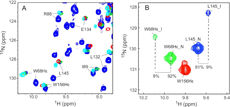
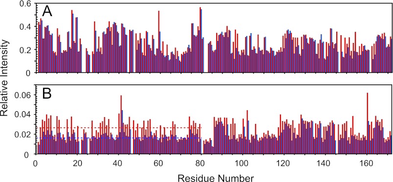
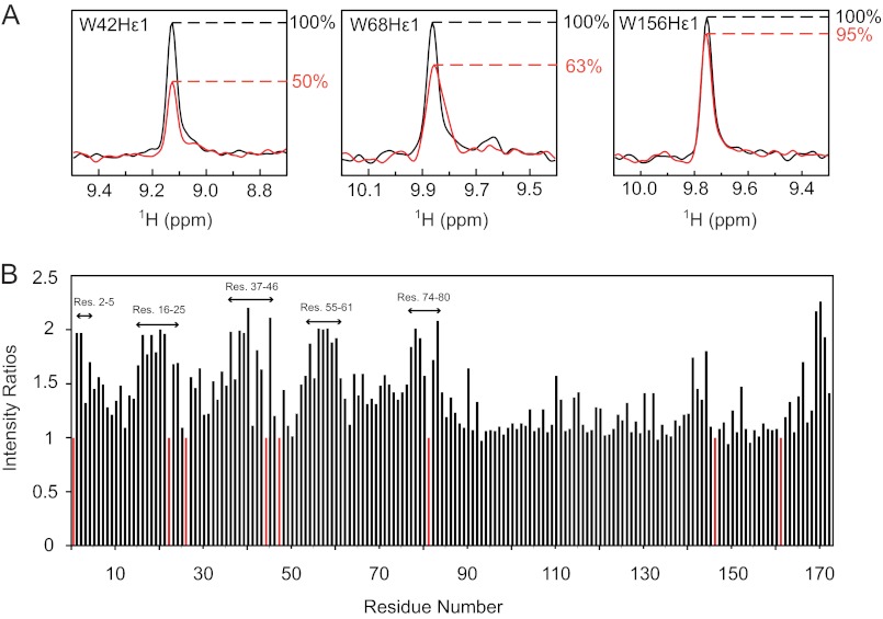
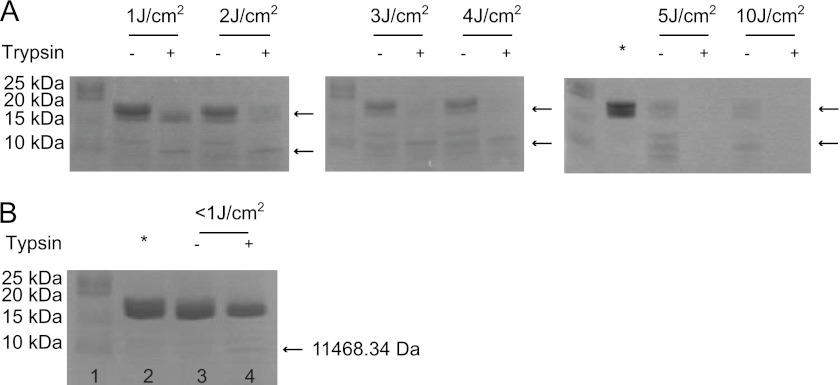
Some mutants of human γD-crystallin are closely linked to congenital cataracts, although the detailed molecular mechanisms of mutant-associated cataract formation are generally not known. Here we report on a recently discovered γD-crystallin mutant (W42R) that has been linked to autosomal dominant, congenital cataracts in a Chinese family. The mutant protein is much less soluble and stable than wild-type γD-crystallin. We solved the crystal structure of W42R at 1.7 Å resolution, which revealed only minor differences from the wild-type structure. Interestingly, the W42R variant is highly susceptible to protease digestion, suggesting the presence of a small population of partially unfolded protein. This partially unfolded species was confirmed and quantified by NMR spectroscopy. Hydrogen/deuterium exchange experiments revealed chemical exchange between the folded and unfolded species. Exposure of wild-type γD-crystallin to UV caused damage to the N-terminal domain of the protein, resulting in very similar proteolytic susceptibility as observed for the W42R mutant. Altogether, our combined data allowed us to propose a model for W42R pathogenesis, with the W42R mutant serving as a mimic for photodamaged γD-crystallin involved in age-related cataract.


Figures

FIGURE 1.
FIGURE 1.
1H-15N HSQC spectra and chemical shift differences between the W42R mutant and HGD. A, superposition of the 1H-15N HSQC spectra of ∼0.1 mm W42R (blue contours) and ∼1 mm HGD (red contours) at 25 °C. 166 of 168 amide resonances were assigned and are labeled with amino acid name and number. HGD amide resonances are labeled in italic. B, combined amide 1H, 15N chemical shift differences between the W42R mutant and HGD versus residue number (Res.). Unassigned resonances are given an arbitrary value of 0.1 ppm.
FIGURE 2.
FIGURE 2.
Crystal structure of human W42R γD-crystallin. A, superposition of the N-td (red, residues 1–81) and C-td (magenta, residues 82–171) coordinates of W42R onto those of full-length HGD (cyan, PDB ID: 1HK0). B, best-fitting the W42R N-td-only coordinates (magenta, residues 1–81) onto full-length HGD (cyan, PDB ID: 1HK0). C, the electron density of residue Arg-42 in W42R, contoured at 1.0 electron density standard deviation. D, contacts around the Arg-42 side chain in the W42R mutant structure (left) and around Trp-42 in the HGD structure (PDB ID: 1HK0, right). Hydrogen bonds are labeled with black dots, and hydrophobic contacts are labeled with black semicircles.
FIGURE 3.
FIGURE 3.
Denaturant induced unfolding of W42R (red) and HGD (blue) by GdnHCl or urea (inset) at 37 °C. All samples contained 10 μg/ml protein in 100 mm sodium phosphate buffer, pH 7.0, 5 mm DTT, 1 mm EDTA and GdnHCl from 0 to 5.5 m and urea from 0 to 3 m. FI, fluorescence intensity ratio.
FIGURE 4.
FIGURE 4.
Superposition of the 1H-15N HSQC spectra of untreated (red) and trypsin-digested (blue) W42R. Trypsin cleavage reactions were performed overnight at 37 °C in 20 mm sodium phosphate, pH 7.0, 5 mm DTT, 0.02% NaN3 with an enzyme-to-substrate ratio of 1:100 (w/w). A comparison between trypsin-digested wild-type HGD and the W42R mutant by SDS-PAGE is shown in the inset. Note that lane 1 contains protein standards, lanes 2 and 3 contain HGD in the absence and presence of trypsin, respectively, and lanes 4 and 5 contain W42R in the absence and presence of trypsin, respectively.
FIGURE 5.
FIGURE 5.
NMR evidence for the presence of a partially (un)folded species of W42R. A, expanded regions of the superposition of the 1H-15N HSQC spectra of W42R, trypsin-digested 15N W42R, HGD C-td, and V75D. Spectra of W42R were recorded at 900 MHz at 37 °C (blue contours), spectra of trypsin-digested 15N W42R were recorded at 800 MHz at 37 °C (green contours), spectra of HGD C-td under native conditions were recorded at 600 MHz at 25 °C (red contours), and spectra of V75D in 3.6 m urea were recorded at 900 MHz at 37 °C (cyan contours), respectively. B, expanded region of the 1H-15N HSQC spectrum of the W42R mutant protein at 900 MHz and 37 °C. The Trp-68 Nϵ1 resonances (green contours), Trp-156 Nϵ1 resonances (red contours), and Leu-145 amide resonances (blue contours) are displayed. Resonances associated with the natively folded protein are labeled with N, and resonances associated with the partially unfolded state are labeled with I.
FIGURE 6.
FIGURE 6.
H/D exchange of HGD and W42R mutant proteins. A, relative intensities of HGD resonances in 50% D2O to HGD resonances in 5% D2O at 0 h (red) and 30 h (blue). B, relative intensities of W42R resonances in 50% D2O to HGD resonances in 5% D2O at 0 h (red) and 30 h (blue). The average relative intensity of resonances in the 1H-15N HSQC spectra the N-td of W42R is indicated by the dotted line.
FIGURE 7.
FIGURE 7.
Effect of UV exposure on 15N HGD. A, slices through the tryptophan Hϵ1 resonances in the 1H-15N HSQC spectra of UV-exposed (red) versus nonexposed (black) protein are superimposed. B, intensity ratios of native resonances of 15N HGD to those of 15N HGD after UV exposure.
FIGURE 8.
FIGURE 8.
UV-C exposure and trypsin digestion of HGD and 15N HGD. A and B, SDS-PAGE of UV-C-exposed and trypsin-digested natural abundance HGD for increasing energy doses (1.0, 2.0, 3.0, 4.0, 5.0 J/cm2) (A) and UV-C-exposed and trypsin-digested 15N HGD (B). In B: lane 1, protein standard; lane 2, 15N HGD; lane 3, 15N HGD incubated at 37 °C overnight without trypsin; lane 4, 15N HGD incubated at 37 °C overnight with trypsin. Nonexposed HGD samples are loaded in the lanes marked by asterisks.

References

    1. Reddy M. A., Bateman O. A., Chakarova C., Ferris J., Berry V., Lomas E., Sarra R., Smith M. A., Moore A. T., Bhattacharya S. S., Slingsby C. (2004) Characterization of the G91del CRYBA1/3-crystallin protein: a cause of human inherited cataract. Hum. Mol. Genet. 13, 945–953 - PubMed
    1. Vijaya R., Gupta R., Panda G., Ravishankar K., Kumaramanickavel G. (1997) Genetic analysis of adult-onset cataract in a city-based ophthalmic hospital. Clin. Genet. 52, 427–431 - PubMed
    1. Rahi J. S., Dezateux C. (2000) Congenital and infantile cataract in the United Kingdom: underlying or associated factors. Invest. Ophthalmol. Vis. Sci. 41, 2108–2114 - PubMed
    1. Lampi K. J., Ma Z., Shih M., Shearer T. R., Smith J. B., Smith D. L., David L. L. (1997) Sequence analysis of βA3, βB3, and βA4 crystallins completes the identification of the major proteins in young human lens. J. Biol. Chem. 272, 2268–2275 - PubMed
    1. Hejtmancik J. F. (2008) Congenital cataracts and their molecular genetics. Semin. Cell Dev. Biol. 19, 134–149 - PMC - PubMed

Publication types

Associated data