. 2012 Dec 14;287(51):42525-32.
doi: 10.1074/jbc.M112.425371. Epub 2012 Nov 2.

cAMP-responsive element modulator α (CREMα) contributes to decreased Notch-1 expression in T cells from patients with active systemic lupus erythematosus (SLE)

Affiliations

cAMP-responsive element modulator α (CREMα) contributes to decreased Notch-1 expression in T cells from patients with active systemic lupus erythematosus (SLE)

Thomas Rauen et al. J Biol Chem. .

Abstract

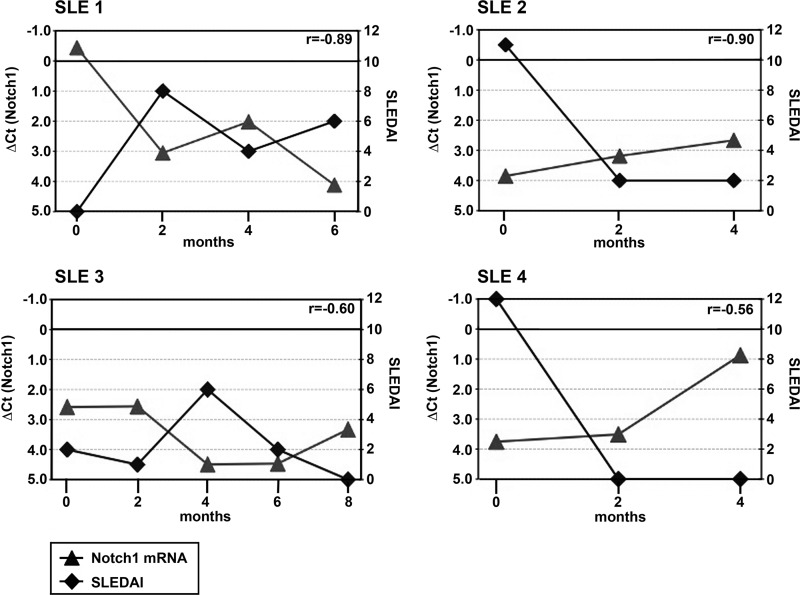
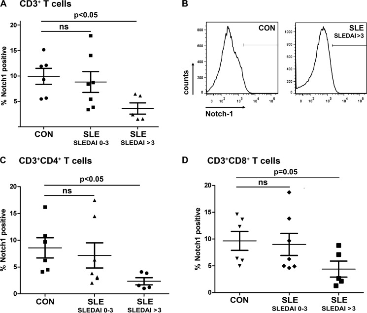
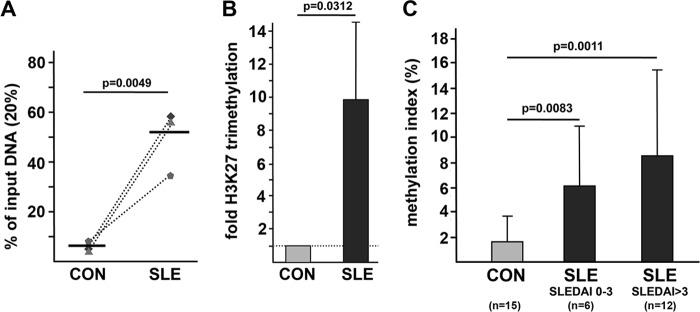
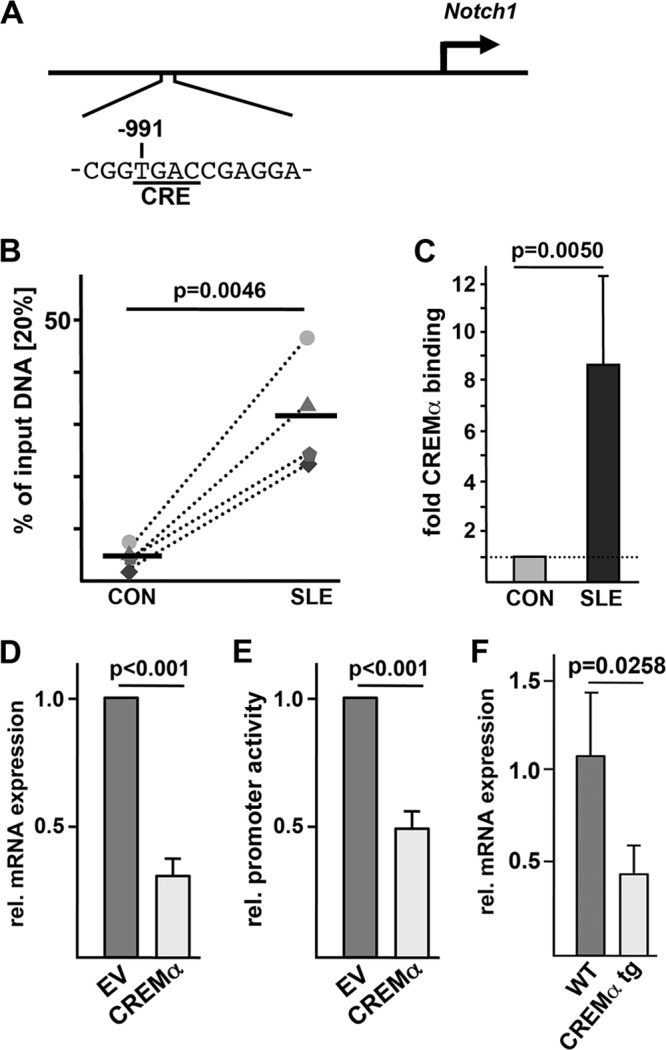
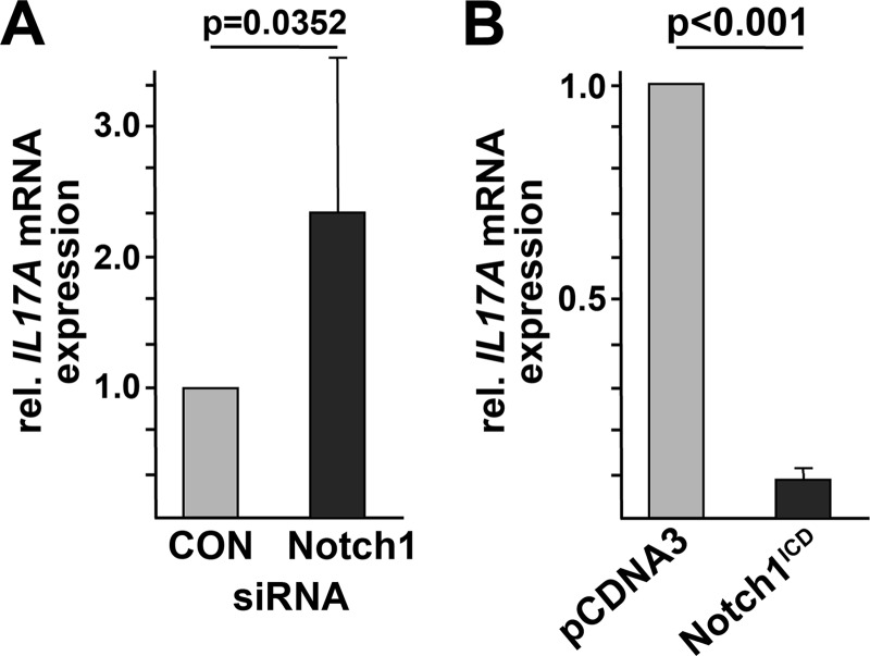
Notch signaling constitutes an evolutionarily conserved pathway that transduces signals between neighboring cells and determines major decisions in cell proliferation, survival, and differentiation. Notch signaling has been shown to play a pivotal role during T cell lineage determination. T lymphocytes from patients with systemic lupus erythematosus (SLE) display a severely altered phenotype with several molecular and functional aberrations, including defective capacities to up-regulate Notch-1 receptor expression upon T cell receptor activation. Here, we demonstrate that basal Notch-1 expression is decreased in T cells from active SLE patients at the mRNA and protein levels in various T cell subpopulations. Notch-1 transcript numbers inversely correlate with disease activity in SLE patients. We provide evidence that both enhanced histone H3 methylation and CpG DNA methylation of the human Notch-1 promoter contribute to decreased Notch-1 expression in SLE T cells. Previous data from our group identified cAMP-responsive element modulator α (CREMα), which is up-regulated in SLE T cells, as a key regulator of epigenetic patterns and gene transcription, e.g. that of IL2 and IL17 genes. In this study, we observed increased CREMα binding to the Notch-1 promoter, which eventually resulted in significantly reduced Notch-1 promoter activity and gene transcription. Notably, decreased Notch-1 levels were associated with elevated IL-17A levels. Our data suggest a role for Notch-1 in SLE immunopathogenesis, and for the first time, we present molecular mechanisms that mediate dysregulated Notch-1 expression in SLE T cells.


Figures

FIGURE 1.
FIGURE 1.
Notch-1 mRNA expression in T cells from SLE patients and healthy controls. Total T cell mRNAs from 24 healthy control individuals (CON) and 61 SLE patients (subgrouped according SLEDAIs) were analyzed for relative Notch-1 expression by real-time qRT-PCR. Crossing points (Ct) were calculated using the second derivative maximum method, an algorithm that requires minimal user input. Normalized Ct values (ΔCt = Ct(target gene) − Ct(average of control genes GAPDH and CD3ϵ)) were used to compare Notch-1 expression levels between different sample groups. To better visualize trends in relative Notch-1 mRNA expression, the y axis is displayed in an inverse manner. Horizontal lines indicate the mean ± S.D. ns, not significant.
FIGURE 2.
FIGURE 2.
Longitudinal analyses of Notch-1 mRNA expression and corresponding SLEDAIs. Total T cell mRNAs from four SLE patients (SLE 1 to SLE 4) were collected every other month, and both relative Notch-1 mRNA expression in total T cells (normalized Ct values; left y axis, ▴) and SLEDAIs (right y axis, ♦) were determined at each time point. Individual Pearson's correlation coefficients (r) are given in the upper right corner of each panel.
FIGURE 3.
FIGURE 3.
Notch-1 protein expression at the surface of T cells from SLE patients and healthy controls. A, CD3+ T cells from healthy control individuals (CON) and SLE patients were analyzed for Notch-1 protein expression by flow cytometry. Percentages of Notch-1+ cells are given in the diagram. B, a representative staining pattern is shown. T helper (CD3+CD4+; C) and cytotoxic (CD3+CD8+; D) T cells were analyzed for Notch-1 protein expression by flow cytometry. Horizontal lines indicate the mean ± S.D. ns, not significant.
FIGURE 4.
FIGURE 4.
Histone methylation and CpG DNA methylation of the Notch-1 promoter are increased in T cells from SLE patients. A, histone H3K27 methylation was analyzed in total T cells from three age-, gender-, and ethnicity-matched control (CON)/SLE pairs by ChIP assays. Dotted lines associate matched samples. A region of interest within the human Notch-1 promoter (harboring a putative CRE) was amplified by qPCR, and the proportion of immunoprecipitated DNA was calculated as relative to the non-immunoprecipitated input DNA in each sample. Subsequently, the ratio of relative expression was calculated between each SLE patient and the corresponding control individual. Horizontal lines represent mean values. B, the dotted line represents the H3K27 methylation status in control T cells, for each of which was set to 1.0. Changes in the methylation status in the matched SLE patient are given in the bar diagram (mean ± S.D.). C, total T cells from 15 healthy controls (light gray bar), 6 inactive SLE patients (dark gray bar), and 12 active SLE patients (black bar) were subjected to methylated DNA immunoprecipitation. The percentage of methylated DNA is given as mean ± S.D.
FIGURE 5.
FIGURE 5.
CREMα binds to and trans-represses the Notch-1 promoter. A, schematic of the human Notch-1 gene promoter indicating the CRE of interest located 991 bp upstream of the start codon. B, ChIP was performed using total T cells from four matched pairs of SLE patients and healthy controls (CON) and anti-CREMα antibody. Immunoprecipitated DNA was analyzed by real-time qRT-PCR using the same primers as used for the data in Fig. 4. Ratios between anti-CREMα antibody-immunoprecipitated and input DNAs are shown. Dotted lines associate data from the matched control/SLE pairs. Horizontal lines represent mean values. C, the percentage of anti-CREMα antibody-immunoprecipitated DNA in T cells from a control individual was set to 1.0, and the relative CREMα binding in the corresponding SLE patient was calculated. Values are given as mean ± S.D. D, pcDNA3 empty vector (EV) or CREMα expression plasmid was transfected into human Jurkat T cells, and 5 h after transfection, mRNA was analyzed for Notch-1 and 18 S rRNA expression by real-time qRT-PCR. E, a 2.1-bp spanning Notch-1 promoter sequence (within luciferase vector pGL3) was transfected into Jurkat T cells in the presence or absence of a CREMα expression plasmid, and 5 h after transfection, the relative (rel.) luciferase activity was determined. F, spleens from FVB WT and CREMα transgenic (tg) mice were collected, and CD4+ T cells were isolated by MACS® sorting. Murine Notch-1 and 18 S rRNA expression was analyzed by real-time qRT-PCR in these cells.
FIGURE 6.
FIGURE 6.
Notch-1 levels control IL-17A synthesis. A, siRNA was used to down-regulate endogenous Notch-1 levels in human Jurkat T cells. (Irrelevant siRNA transfection was used in the control (CON) assays.) mRNA from these cells was analyzed for relative (rel.) IL17A expression by real-time qRT-PCR. B, human Jurkat T cells were transfected with a constitutively active Notch-1 construct (i.e. the intracellular receptor domains (Notch1ICD)) or pcDNA3 empty vector. Cells were harvested 5 h after transfection, and relative IL17A expression was determined by real-time qRT-PCR.

References

    1. Tsokos G. C. (2011) Systemic lupus erythematosus. N. Engl. J. Med. 365, 2110–2121 - PubMed
    1. Grammatikos A. P., Tsokos G. C. (2012) Immunodeficiency and autoimmunity: lessons from systemic lupus erythematosus. Trends Mol. Med. 18, 101–108 - PMC - PubMed
    1. Ahlmann M., Varga G., Sturm K., Lippe R., Benedyk K., Viemann D., Scholzen T., Ehrchen J., Müller F. U., Seidl M., Matus M., Tsokos G. C., Roth J., Tenbrock K. (2009) The cyclic AMP response element modulator α suppresses CD86 expression and APC function. J. Immunol. 182, 4167–4174 - PMC - PubMed
    1. Ghosh D., Kis-Toth K., Juang Y. T., Tsokos G. C. (2012) CREMα suppresses spleen tyrosine kinase expression in normal but not systemic lupus erythematosus T cells. Arthritis Rheum. 64, 799–807 - PMC - PubMed
    1. Rauen T., Hedrich C. M., Juang Y. T., Tenbrock K., Tsokos G. C. (2011) cAMP-responsive element modulator (CREM) α protein induces interleukin-17A expression and mediates epigenetic alterations at the interleukin-17A gene locus in patients with systemic lupus erythematosus. J. Biol. Chem. 286, 43437–43446 - PMC - PubMed

Publication types

MeSH terms

LinkOut - more resources