. 2013 Apr;229(5):645-59.
doi: 10.1002/path.4125.

Proximal tubular cells contain a phenotypically distinct, scattered cell population involved in tubular regeneration

Affiliations

Proximal tubular cells contain a phenotypically distinct, scattered cell population involved in tubular regeneration

Bart Smeets et al. J Pathol. 2013 Apr.

Abstract

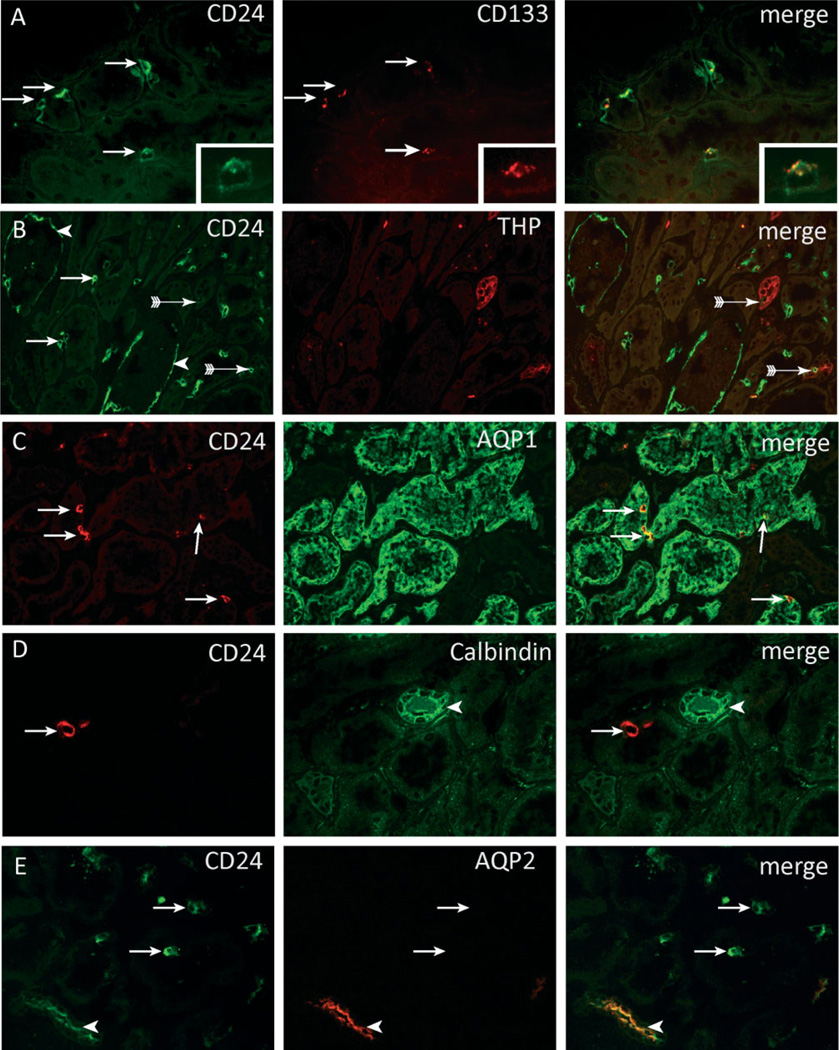
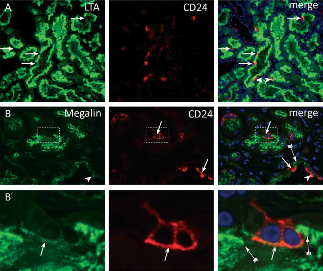
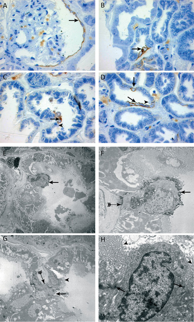
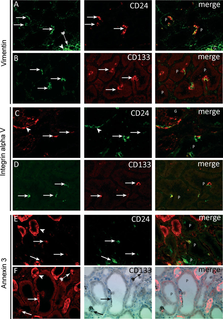
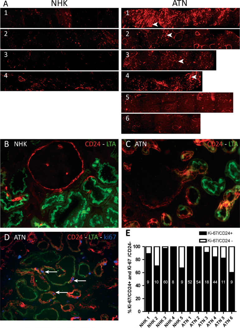
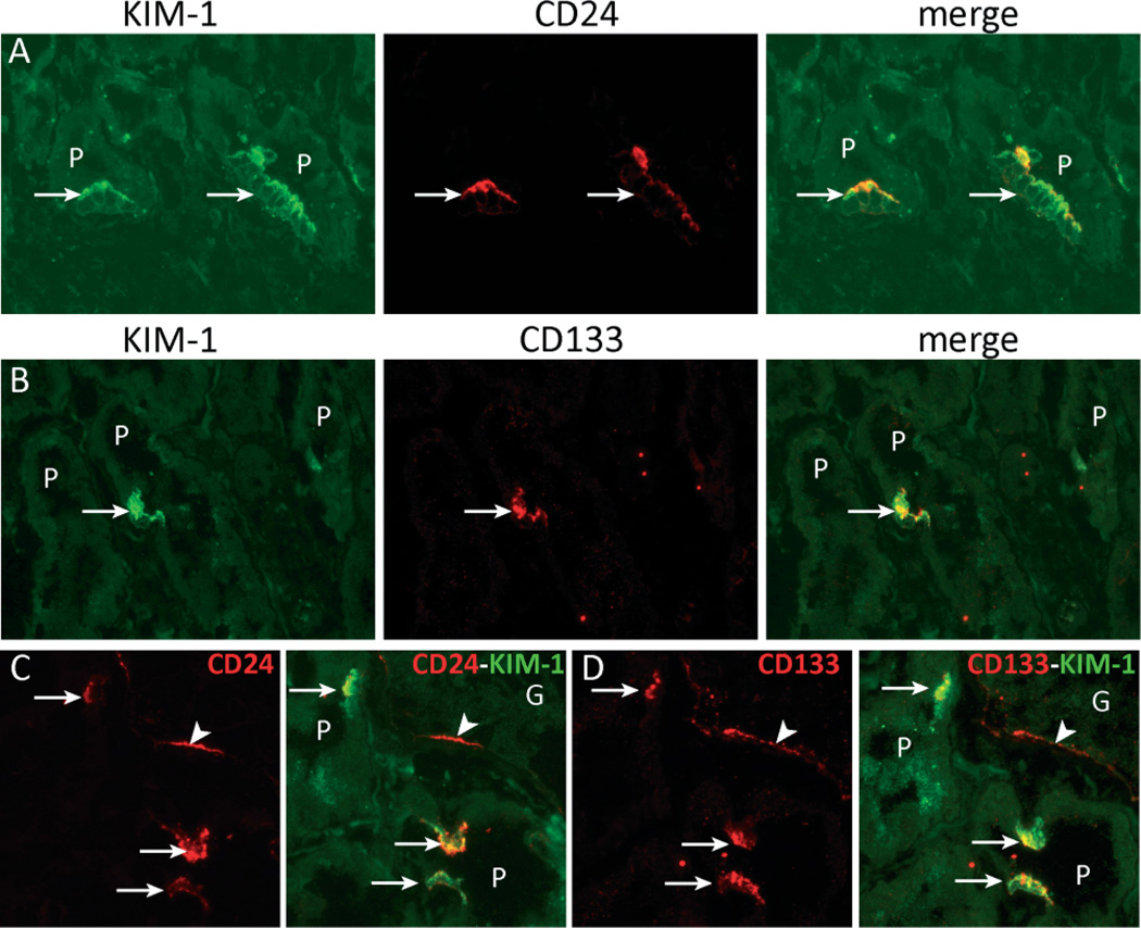
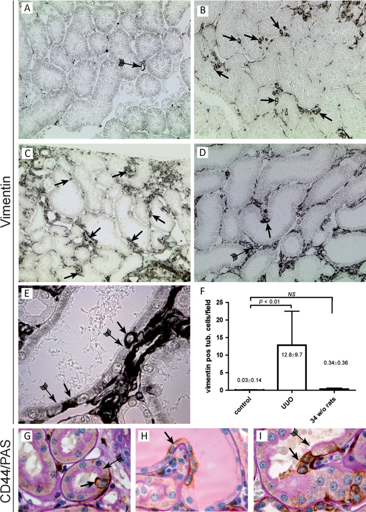
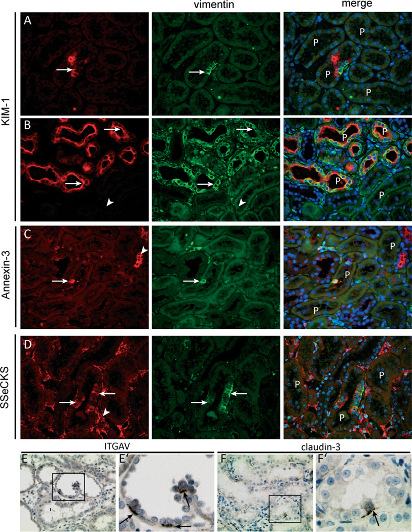
Regeneration of injured tubular cells occurs after acute tubular necrosis primarily from intrinsic renal cells. This may occur from a pre-existing intratubular stem/progenitor cell population or from any surviving proximal tubular cell. In this study, we characterize a CD24-, CD133-, and vimentin-positive subpopulation of cells scattered throughout the proximal tubule in normal human kidney. Compared to adjacent 'normal' proximal tubular cells, these CD24-positive cells contained less cytoplasm, fewer mitochondria, and no brush border. In addition, 49 marker proteins are described that are expressed within the proximal tubules in a similar scattered pattern. For eight of these markers, we confirmed co-localization with CD24. In human biopsies of patients with acute tubular necrosis (ATN), the number of CD24-positive tubular cells was increased. In both normal human kidneys and the ATN biopsies, around 85% of proliferating cells were CD24-positive - indicating that this cell population participates in tubular regeneration. In healthy rat kidneys, the novel cell subpopulation was absent. However, upon unilateral ureteral obstruction (UUO), the novel cell population was detected in significant amounts in the injured kidney. In summary, in human renal biopsies, the CD24-positive cells represent tubular cells with a deviant phenotype, characterized by a distinct morphology and marker expression. After acute tubular injury, these cells become more numerous. In healthy rat kidneys, these cells are not detectable, whereas after UUO, they appeared de novo - arguing against the notion that these cells represent a pre-existing progenitor cell population. Our data indicate rather that these cells represent transiently dedifferentiated tubular cells involved in regeneration.


Conflict of interest statement

No conflicts of interest were declared.

Figures

Figure 1
Figure 1
Localization of CD24- and CD133-positive cells within the kidney. (A – E) Within normal human kidney, CD24-positive cells were scattered as single cells or in small groups of 2 – 4 cells throughout the kidney cortex (arrows). The CD24-positive cells in proximal tubuli co-expressed the cell surface marker CD133 (A, arrows). The insets show a higher magnification of a CD24- and CD133-positive cell. CD24 is expressed on both the basal and the apical membrane. CD133 is expressed on the apical membrane. CD24 expression was also observed in parietal cells (PECs) on Bowman’s capsule (B, arrowheads). Co-staining with CD24 and THP showed rare cells that expressed both markers (B, arrows with tail). The majority of the CD24-positive cells were present in aquaporin-1-positive proximal tubular structures (C, arrows). No co-localization was observed between calbindin (D, arrowhead) and CD24. Aquaporin-2-positive collecting ducts cells showed apical staining for CD24 (E, arrowhead).
Figure 2
Figure 2
CD24-positive cells lack a brush border. Double staining for (A) Lotus tetragonolobus lectin (LTA) and (B) megalin showed that most CD24-positive cells lacked expression of these brush border molecules (arrows). (B’) Magnifications of panel B showing that the CD24-positive cells show no or weak staining for megalin. Neighbouring CD24-negative cells were megalin-positive (B, B’, arrow with tail). A small number of CD24-positive cells showed a normal LTA or megalin-positive brush border (A, B, arrowheads).
Figure 3
Figure 3
Ultrastructural analysis of CD24 immunolabelled cells in semi-thin and ultrathin sections. (A – D) CD24 (brown staining) is expressed by the parietal epithelial cells of Bowman’s capsule (A, arrow) and a few proximal tubular cells (B – D, arrows). An abrupt termination of the brush border was visible on cells adjacent to the CD24-positive cells (C, D, arrowheads). Immunoelectron (E – G) and transmission electron microscopy (H). The morphology of CD24-positive cells (black outlining, E – G, arrows) differed from that of normal proximal tubular cells. CD24-positive cells contained less cytoplasm, fewer mitochondria, and the brush border was mostly rudimentary or absent compared with the brush border of adjacent CD24-negative cells (G, H, arrowheads). Of note, some CD24-positive cells contained a brush border. Panel G shows two CD24-positive cells adjacent to each other, one of which lacks a brush border (G, arrow) and the other does not (G, arrowhead). In addition, the basolateral membrane lacked the typical invaginations (F, G, arrow with tail). Electron microscopy revealed desmosomes between the two morphologically different proximal tubular cells (H, arrowheads).
Figure 4
Figure 4
CD24- and CD133-positive cells exhibit distinct marker expression. In contrast to neighbouring cells, CD24- and CD133-positive proximal tubular cells (A – F, arrows) show expression of vimentin (A, B), integrin αV (C, D), and annexin 3 (E, F). Vimentin (A, B) was strongly expressed by podocytes (A, arrowhead) and parietal epithelial cells (A, arrow with tail) but also showed co-localization in the CD24- and CD133-positive proximal tubular cells (A, B, arrows, respectively). Integrin αV (C, D) was expressed by the parietal cells (C, arrowhead) and the CD24- and CD133-positive proximal tubular cells (C, D, arrows, respectively). Annexin 3 (E, F) was expressed in distal tubuli (arrowhead) and in the CD24- and CD133-positive proximal tubular cells (arrows). The arrow with tail in panel F shows an annexin 3 cell negative for CD133. G = glomerulus; P = proximal tubules.
Figure 5
Figure 5
Increased number of CD24-positive cells in acute tubular necrosis. (A) Comparison of microscopic scans of whole biopsies of normal human kidneys (NHK) with biopsies of acute tubular necrosis (ATN) showed that the number of CD24-positive tubular structures was increased in most (1 – 4) of the ATN biopsies. The low CD24 expression in 5 – 6 correlated with the relative mild necrosis visible in the PAS staining of these biopsies and the relatively low serum creatinine levels. The increased number of CD24-positive cells in ATN (C) compared with NHK (B) correlated with a high number of proliferating cells in the ATN biopsies (D, Ki-67-positive cell). (D) Triple staining of CD24, LTA, and Ki-67 showed that Ki-67 was expressed predominantly in CD24-positive proximal tubular cells (arrows). (E) Quantitative Ki-67, CD24, and LTA triple staining. The bars show the relative percentage of Ki-67-positive cells that are either CD24-positive (black bar) or CD24-negative (grey bar). The vast majority (~70 – 100%) of proliferating cells in both NHK and ATN biopsies were CD24-positive. Data labels: number of Ki-67 cells analysed in each biopsy.
Figure 6
Figure 6
CD24- and CD133-positive proximal tubular cells express kidney injury molecule-1 (KIM-1). (A – D) Double immunofluorescence staining showing co-localization between CD24 or CD133 and KIM-1 in the CD24- and CD133-positive proximal tubule cell population (arrows). Panels C and D are serial sections stained for CD24/KIM-1 and CD133/KIM-1, respectively. CD24 and CD133 are expressed by the cells of Bowman’s capsule (arrowhead) and by the same KIM-1-positive proximal tubule cells (arrows).
Figure 7
Figure 7
Detection of scattered vimentin- and CD44-positive tubular cells in the unilateral ureteral obstruction model (UUO). (A) Vimentin expression in the contralateral control kidney. Vimentin is exclusively expressed in the peritubular capillaries (10× magnification). (B – D) In kidneys that underwent UUO, de novo expression of vimentin was observed in proximal tubular cells. The cells were frequently scattered single cells or small groups of cells (arrows). Also in the interstitium, the expression of vimentin was increased (D, arrow with tail). (B) 20×, (C) 10×, and (D) 20× magnification. (E) Higher magnification (60×) of a proximal tubule showing scattered vimentin-positive cells with an undeveloped brush border (arrows) juxtaposed to differentiated proximal tubular cells with a normal brush border (arrows with tail). (F) Quantitative analysis of the number of vimentin-positive tubular cells per microscopic field (20× magnification) in the contralateral and UUO kidneys of 7-week-old rats and in the kidneys of aged 34-week-old rats. (G – I) CD44/PAS co-staining shows single or small groups of CD44-positive tubular cells with an undeveloped brush border (arrows) juxtaposed to differentiated proximal tubular cells with a normal brush border (arrows with tail) (60× magnification).
Figure 8
Figure 8
Vimentin-positive proximal tubular cells exhibit distinct marker expression. (A, B) The vimentin-positive cells in the injured rat kidneys co-expressed KIM-1. Vimentin and Kim-1 co-expression was seen in single cells or small groups of cells (A, arrow) but also in complete proximal tubular structures in segments of the kidney with more advanced tubular injury (B, arrows). Adjacent normal-appearing tubuli were mostly negative for both markers (B, arrowhead). (C) Within proximal tubuli, vimentin co-localized with annexin 3 (arrow). Annexin 3 was also expressed in other tubular structures (arrowhead). (D) Vimentin-positive proximal tubular cells co-expressed SSeCKS (arrows), which was also expressed by endothelial cells (arrowhead) and parietal cells (not shown). (E, F) Within the affected rat kidneys, both integrin αV and claudin-3 were expressed by a few proximal tubuli in a scattered pattern. E’ and F’ show a zoomed picture of the insets in E and F, respectively, and show mostly basolateral staining of both markers in a subset of the proximal tubular cells.

Comment in

References

    1. Vogetseder A, Palan T, Bacic D, et al. Proximal tubular epithelial cells are generated by division of differentiated cells in the healthy kidney. Am J Physiol Cell Physiol. 2007;292:C807–C813. - PubMed
    1. Vogetseder A, Picard N, Gaspert A, et al. Proliferation capacity of the renal proximal tubule involves the bulk of differentiated epithelial cells. Am J Physiol Cell Physiol. 2008;294:C22–C28. - PubMed
    1. Humphreys D, Valerius TM, Kobayashi A, et al. Intrinsic epithelial cells repair the kidney after injury. Cell Stem Cell. 2008;2:284–291. - PubMed
    1. Song J, Czerniak S, Wang T, et al. Characterization and fate of telomerase-expressing epithelia during kidney repair. J Am Soc Nephrol. 2011;22:2256–2265. - PMC - PubMed
    1. Humphreys BD, Czerniak S, DiRocco DP, et al. Repair of injured proximal tubule does not involve specialized progenitors. Proc Natl Acad Sci U S A. 2011;108:9226–9231. - PMC - PubMed

Publication types

MeSH terms