. 2013 May 15;132(10):2316-26.
doi: 10.1002/ijc.27924. Epub 2012 Nov 26.

Cancer cells preferentially lose small chromosomes

Affiliations

Cancer cells preferentially lose small chromosomes

Pascal H G Duijf et al. Int J Cancer. .

Abstract

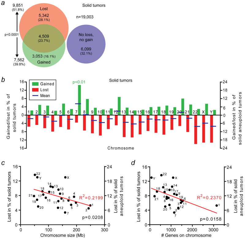
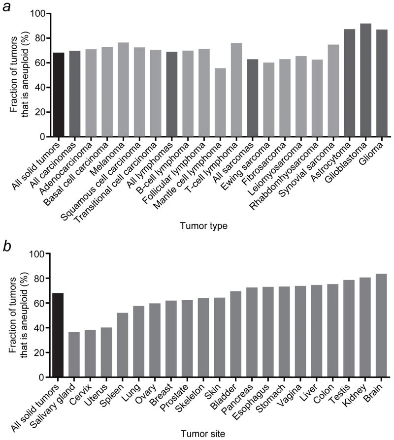
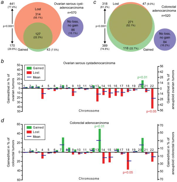
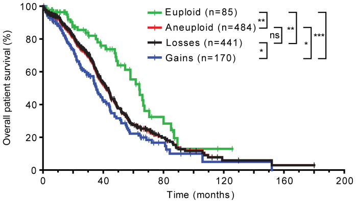
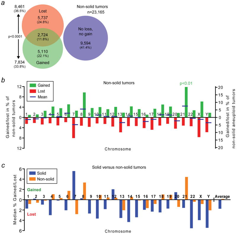
Genetic and genomic aberrations are the primary cause of cancer. Chromosome missegregation leads to aneuploidy and provides cancer cells with a mechanism to lose tumor suppressor loci and gain extra copies of oncogenes. Using cytogenetic and array-based comparative genomic hybridization data, we analyzed numerical chromosome aneuploidy in 43,205 human tumors and found that 68% of solid tumors are aneuploid. In solid tumors, almost all chromosomes are more frequently lost than gained with chromosomes 7, 12 and 20 being the only exceptions with more frequent gains. Strikingly, small chromosomes are lost more readily than large ones, but no such inverse size correlation is observed with chromosome gains. Because of increasing levels of proteotoxic stress, chromosome gains have been shown to slow cell proliferation in a manner proportional to the number of extra gene copies gained. However, we find that the extra chromosome in trisomic tumors does not preferentially have a low gene copy number, suggesting that a proteotoxicity-mediated proliferation barrier is not sustained during tumor progression. Paradoxically, despite a bias toward chromosome loss, gains of chromosomes are a poor prognostic marker in ovarian adenocarcinomas. In addition, we find that solid and non-solid cancers have markedly distinct whole-chromosome aneuploidy signatures, which may underlie their fundamentally different etiologies. Finally, preferential chromosome loss is observed in both early and late stages of astrocytoma. Our results open up new avenues of enquiry into the role and nature of whole-chromosome aneuploidy in human tumors and will redirect modeling and genetic targeting efforts in patients.


Figures

Figure 1
Figure 1
Human solid tumors preferentially lose small chromosomes. (a) Area-proportional Venn diagram indicating the numbers and fractions of a total of 19,003 human solid tumors that have lost, gained, concomitantly lost and gained or have neither lost nor gained whole-chromosomes. (b) Rates at which human solid tumors gain or lose individual whole-chromosomes. The blue bars indicate the means of the gain and loss rates. Chromosome 7 is gained significantly more frequently than any of the other chromosomes (p<0.01; two-sided Grubbs’ test). (c) Scatter plot of chromosome size in megabases (Mb) in relationship to the rate at which the chromosome is lost in human solid tumors. (d) Scatter plot of the number of genes on the chromosome in relationship to the rate at which the chromosome is lost in human solid tumors.
Figure 2
Figure 2
Whole-chromosome aneuploidy rates in human solid tumors vary among tumor types and tumor sites. (a) Whole-chromosome aneuploidy rates for selected tumor types. (b) Whole-chromosome aneuploidy rates for tumors that developed in various different organs.
Figure 3
Figure 3
Different types of human solid tumors have distinct whole-chromosome aneuploidy signatures. (a) Area-proportional Venn diagram as in Figure 1a. However, these data are derived from The Cancer Genome Atlas (TCGA) array-based comparative genomic hybridization (aCGH) data of 570 human ovarian serous cystadenocarcinomas. (b) Rates at which ovarian serous cystadenocarcinomas gain or lose individual whole-chromosomes. Blue bars indicate the means of the gain and loss rates. Chromosome 20 is gained significantly more frequently than any of the other chromosomes (p<0.01; two-sided Grubbs’ test), whereas chromosome 22 is significantly more frequently lost than any of the other chromosomes (p<0.05; two-sided Grubbs’ test). (c) Area-proportional Venn diagram of 520 human colorectal adenocarcinomas using aCHG-derived data from TCGA. The areas between (a) and (c) are not proportional. (d) Individual whole-chromosome gain and loss rates as in (b) but for colorectal adenocarcinomas. Chromosomes 13 and 18 are more frequently gained and lost, respectively, than the other chromosomes (p<0.01 and p<0.05, respectively; two-sided Grubbs’ tests).
Figure 4
Figure 4
Whole-chromosome gains are more malignant than whole-chromosome losses in human ovarian cystadenocarcinoma. The overall survival of 569 ovarian serous cystadenocarcinoma patients is plotted. All p values are calculated using the log-rank test and summarized as follows: ns, not significant (i.e., p>0.05); *, 0.01
Figure 5
Figure 5
Human solid and non-solid tumors have markedly different whole-chromosome aneuploidy signatures. (a) Area-proportional Venn diagram as in Figure 1a but for non-solid tumors. (b) Whole-chromosome gain and loss rates for individual chromosomes in non-solid cancers. The blue bars indicate the mean of the gain and loss rates. Chromosome 21 is gained significantly more frequently than any of the other chromosomes (p<0.01; two-sided Grubbs’ test). (c) Comparison of the means of the gain and loss rates of each chromosome for solid (blue) and non-solid tumors (orange). These frequencies represent the respective blue bars in Figures 1b and 5b. Bars above the x-axis indicate a bias towards gain, those below the axis a bias towards loss. The average on the far right corresponds to the average of the plotted gain and negative loss rates of all chromosomes. See also main text for details.
Figure 6
Figure 6
A comparison between early- and late-stage whole-chromosome aneuploidies in human astrocytoma provides insights into tumor evolution. (a) Summary of the most striking differences in whole-chromosome aneuploidy signatures between stage I–II and stage III–IV astrocytomas (see also main text and Supporting Information Figure 4). (b) The number of whole-chromosome aberrations per tumor increases during tumor progression.

References

    1. Albertson DG, Collins C, McCormick F, Gray JW. Chromosome aberrations in solid tumors. Nat Genet. 2003;34:369–76. - PubMed
    1. Carter SL, Eklund AC, Kohane IS, Harris LN, Szallasi Z. A signature of chromosomal instability inferred from gene expression profiles predicts clinical outcome in multiple human cancers. Nat Genet. 2006;38:1043–8. - PubMed
    1. McClelland SE, Burrell RA, Swanton C. Chromosomal instability: a composite phenotype that influences sensitivity to chemotherapy. Cell Cycle. 2009;8:3262–6. - PubMed
    1. Sotillo R, Schvartzman JM, Socci ND, Benezra R. Mad2-induced chromosome instability leads to lung tumour relapse after oncogene withdrawal. Nature. 2010;464:436–40. - PMC - PubMed
    1. Guerrero AA, Gamero MC, Trachana V, Futterer A, Pacios-Bras C, Diaz-Concha NP, Cigudosa JC, Martinez AC, van Wely KH. Centromere-localized breaks indicate the generation of DNA damage by the mitotic spindle. Proc Natl Acad Sci U S A. 2010;107:4159–64. - PMC - PubMed

Publication types