. 2013 Feb;65(2):519-28.
doi: 10.1002/art.37763.

Association of fibromyalgia with altered skeletal muscle characteristics which may contribute to postexertional fatigue in postmenopausal women

Affiliations

Association of fibromyalgia with altered skeletal muscle characteristics which may contribute to postexertional fatigue in postmenopausal women

Ratchakrit Srikuea et al. Arthritis Rheum. 2013 Feb.

Abstract

Objective: To identify muscle physiologic properties that may contribute to postexertional fatigue and malaise in women with fibromyalgia (FM).

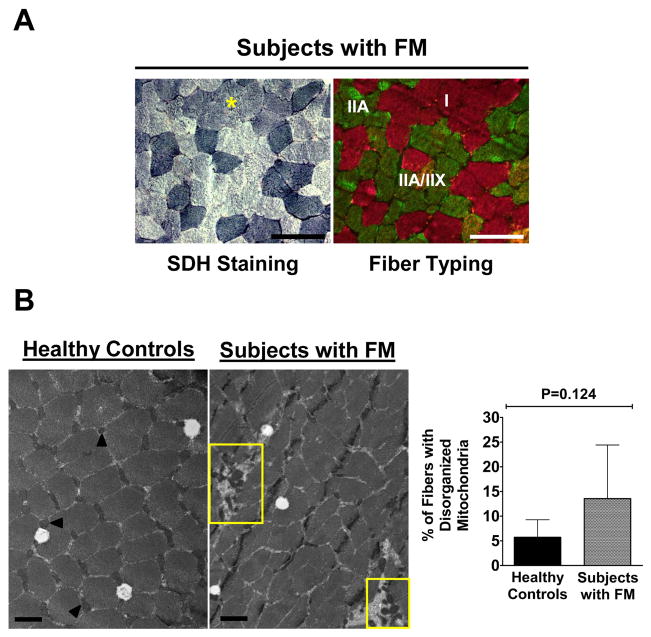
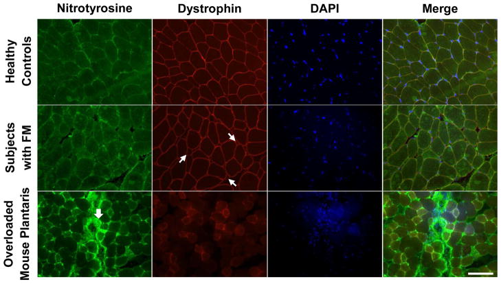
Methods: Healthy postmenopausal women with (n = 11) and without (n = 11) FM, ages 51-70 years, participated in this study. Physical characteristics and responses to self-reported questionnaires were evaluated. Strength loss and tissue oxygenation in response to a fatiguing exercise protocol were used to quantify fatigability and the local muscle hemodynamic profile. Muscle biopsies were performed to assess between-group differences in baseline muscle properties using histochemical, immunohistochemical, and electron microscopic analyses.

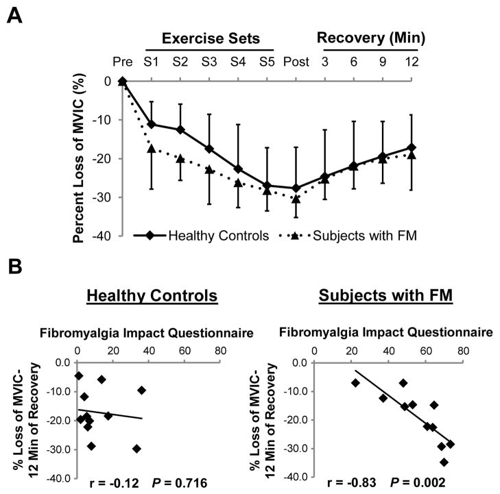
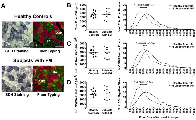
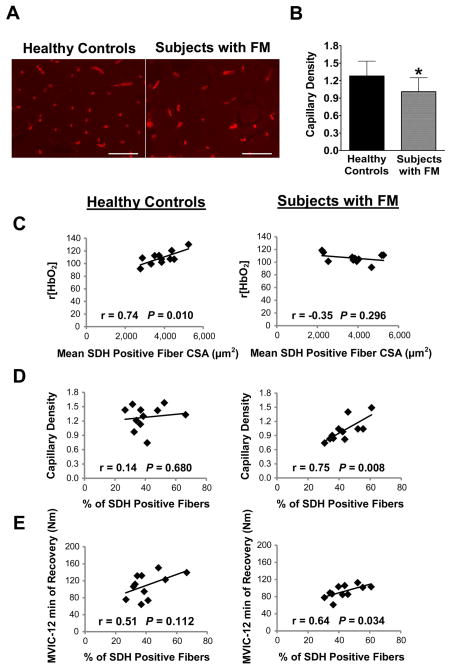
Results: There was no significant difference between healthy controls and FM patients in muscle fatigue in response to exercise. However, self-reported fatigue and pain were correlated with prolonged loss of strength following 12 minutes of recovery in patients with FM. Although there was no difference in percent succinate dehydrogenase (SDH)-positive (type I) and SDH-negative (type II) fibers or in mean fiber cross-sectional area between groups, FM patients exhibited greater variability in fiber size and altered fiber size distribution. In healthy controls only, fatigue resistance was strongly correlated with the size of SDH-positive fibers and hemoglobin oxygenation. In contrast, FM patients with the highest percentage of SDH-positive fibers recovered strength most effectively, and this was correlated with capillary density. However, overall, capillary density was lower in the FM group.

Conclusion: Peripheral mechanisms, i.e., altered muscle fiber size distribution and decreased capillary density, may contribute to postexertional fatigue in FM. Understanding of these defects in fibromyalgic muscle may provide valuable insight with regard to treatment.


Figures

Figure 1
Figure 1. Percent loss in MVIC following the fatiguing exercise protocol is correlated with self-reported assessment in subjects with FM
(A) Maximum voluntary isometric contractions (MVICs) were recorded and plotted against time periods (pre-exercise, after each exercise set (S1–5), and during recovery. (B) Correlation between fibromyalgia impact questionnaire score and % loss of MVIC following 12-min of recovery from fatiguing exercise was observed in subjects with FM but not healthy controls.
Figure 2
Figure 2. Defects in fiber size distribution and loss of correlation to fatigue in subjects with FM
(A) SDH positive fibers (blue) were strongly correlated with type I fibers (pink fibers in right panels) in both healthy controls and subjects with FM. Rarely, type IIA fibers (green in right panel of healthy control) were SDH positive (asterisks). Most IIX fibers also expressed IIA myosin heavy chain (orange staining in right panels) in both groups. Scale bars = 100 μm. Mean fiber cross-sectional area (CSA) and size distribution of total (B); SDH positive (C) and SDH negative (D) fibers. The distribution of fiber sizes differed significantly as determined by the Komogorov-Smirnov (K-S) test, with more small fibers present in subjects with FM. A total of 3,737 fibers for healthy controls (1,425 SDH positive fibers and 2,312 SDH negative fibers) and 3,556 fibers for subjects with FM (1,498 SDH positive fibers and 2,058 SDH negative fibers) were measured for CSA quantification.
Figure 3
Figure 3. Weak SDH positive fiber staining in type I fibers in FM muscle and mitochondrial distribution
(A) Representative weak SDH positive fibers (asterisk, left panel) in a cluster of type I fibers (pink fibers, right panel) in subjects with FM. Images were captured at ×200, scale bars = 100 μm. (B) Representative electron micrographs (EM) showing normal interfibrillar mitochondria in a healthy control (arrows, left panel), compared to disrupted organization in a subject with FM (yellow rectangles). Images were captured at ×13,000, scale bars = 1 μm. Bar graph represents quantification of the frequency of fibers with disorganized mitochondria. Values are means ± SD (n=6/group).
Figure 4
Figure 4. Reduced capillary density and altered correlations with tissue oxygenation, oxidative fibers and strength recovery in FM muscle
(A) Capillaries were identified by lectin staining (red). Scale bars = 100 μm. (B) A significant decrease in capillaries per fiber in subjects with FM compared to healthy controls was detected. * indicates P < 0.05, values are means ± SD (n=11/group). (C) Correlation between r[HbO2] and mean SDH positive fiber CSA was observed in healthy controls but not in subjects with FM. (D) A correlation between capillary density and % SDH positive fibers was observed in subjects with FM but not in healthy controls. (E) A weak correlation between strength following 12-min of recovery, measured by maximum voluntary isometric contractions (MVICs), and % SDH positive fibers was observed in subjects with FM.
Figure 5
Figure 5. No apparent muscle morphologic defects in subjects with FM
Anti-nitrotyrosine (green) and anti-dystrophin (red) antibodies, together with DAPI staining (blue) were used to visualize muscle fiber structure and nuclei. Damaged mouse muscle from overloading was used as a positive control for nitrotyrosine staining. Small arrows represent small fibers in fibromyalgic muscle and large arrow denotes nitrotyrosine accumulation in overloaded mouse plantaris muscle. Scale bar = 100 μm.

References

    1. Abeles AM, Pillinger MH, Solitar BM, Abeles M. Narrative review: the pathophysiology of fibromyalgia. Ann Intern Med. 2007;146:726–34. - PubMed
    1. Cho HJ, Skowera A, Cleare A, Wessely S. Chronic fatigue syndrome: an update focusing on phenomenology and pathophysiology. Curr Opin Psychiatry. 2006;19:67–73. - PubMed
    1. Mease P, Arnold LM, Bennett R, Boonen A, Buskila D, Carville S, et al. Fibromyalgia syndrome. J Rheumatol. 2007;34:1415–25. - PubMed
    1. Wolfe F, Ross K, Anderson J, Russell IJ, Hebert L. The prevalence and characteristics of fibromyalgia in the general population. Arthritis Rheum. 1995;38:19–28. - PubMed
    1. Yunus MB. Gender differences in fibromyalgia and other related syndromes. J Gend Specif Med. 2002;5:42–7. - PubMed

Publication types