. 2012 Nov 1;4(11):a011338.
doi: 10.1101/cshperspect.a011338.

Mitochondrial quality control mediated by PINK1 and Parkin: links to parkinsonism

Affiliations

Mitochondrial quality control mediated by PINK1 and Parkin: links to parkinsonism

Derek Narendra et al. Cold Spring Harb Perspect Biol. .

Abstract

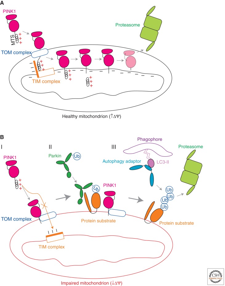
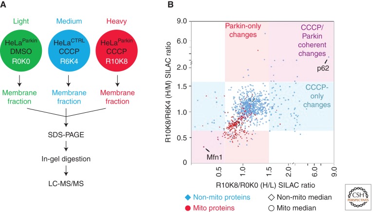
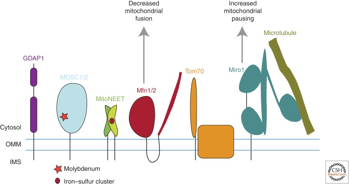
Mutations in Parkin or PINK1 are the most common cause of recessive familial parkinsonism. Recent studies suggest that PINK1 and Parkin form a mitochondria quality control pathway that identifies dysfunctional mitochondria, isolates them from the mitochondrial network, and promotes their degradation by autophagy. In this pathway the mitochondrial kinase PINK1 senses mitochondrial fidelity and recruits Parkin selectively to mitochondria that lose membrane potential. Parkin, an E3 ligase, subsequently ubiquitinates outer mitochondrial membrane proteins, notably the mitofusins and Miro, and induces autophagic elimination of the impaired organelles. Here we review the recent rapid progress in understanding the molecular mechanisms of PINK1- and Parkin-mediated mitophagy and the identification of Parkin substrates suggesting how mitochondrial fission and trafficking are involved. We also discuss how defects in mitophagy may be linked to Parkinson's disease.


Figures

Figure 1.
Figure 1.
Regulation of the PINK1-Parkin mitochondrial quality control pathway by inner mitochondrial membrane potential. (A) In healthy mitochondria, PINK1 is imported to the inner mitochondrial membrane by the TOM (translocase of the outer membrane) and TIM23 (translocase of the inner membrane 23) complexes, along a path that depends on the voltage component of the mitochondrial inner membrane potential (ΔΨ). At the inner membrane, the mitochondrial-targeting signal (MTS) of PINK1 is cleaved by the matrix metalloprotease. PINK1 is cleaved again in its transmembrane domain by the rhomboid protease PARL. The resulting PINK1 cleavage product is unstable and is degraded in a manner that depends on proteasomal activity. (B, I) In impaired mitochondria with collapsed ΔΨ, PINK1 cannot be imported to the inner membrane along the ΔΨ-dependent TIM23 pathway. Instead, PINK1 is directed to the outer mitochondrial membrane by a cryptic signal amino terminal to its transmembrane domain, in which it associates with the TOM complex. (B, II) The accumulation of PINK1 on the outer mitochondrial membrane recruits Parkin from the cytosol and activates its ubiquitin ligase activity. Parkin ubiquitinates outer membrane proteins preferentially on the mitochondrion on which PINK1 has accumulated. (B, III) Ubiquitination of outer mitochondrial membrane proteins by Parkin leads either to their degradation by the proteasome or to the recruitment of ubiquitin-binding adaptor proteins to effect the removal of the damaged mitochondrion by autophagy.
Figure 2.
Figure 2.
Quantitative proteomics-based screen for the identification of Parkin substrates and adaptors in the PINK1/Parkin pathway. (A) Schematic depicting the experimental setup. Three cell lines were labeled with stable isotopes of arginine and lysine to give a light (L) labeled HeLa cell line stably expressing YFP-Parkin treated with vehicle (HeLaParkin); a medium (M) labeled HeLa cell line without Parkin expression treated with the mitochondrial uncoupler CCCP for 2 h (HeLaCTRL); and a heavy (H) labeled HeLaParkin cell line treated with CCCP. A crude membrane fraction was extracted from each cell line by permeabilizing the membranes with a low dose of digitonin, isolating the membranes by centrifugation, and then extracting proteins from the remaining membranes with DDM (n-dodecyl-β-maltoside). The protein extracts from each cell line were mixed in a 1:1:1 ratio, before separation on a SDS-PAGE gel, in-gel digestion, and LC-MS/MS on an Orbitrap instrument with a top 5 duty cycle. The data was analyzed in MaxQuant. (B) Scatter plot depicting H/L (x-axis) and H/M (y-axis) ratios for 992 proteins identified in the experiment. Proteins with “mitochondrial” in the protein name appear as red circles, whereas the others appear as blue diamonds. The median for proteins annotated as mitochondrial is shifted down and to the left relative to the median for other proteins consistent with the en masse degradation of mitochondrial proteins following treatment with CCCP in the presence of Parkin expression. Proteins in the upper right quadrant represent potential adaptors recruited to mitochondria. Proteins in the lower left quadrant represent potential substrates of Parkin. The protein with the largest increase along the x- and y-axes, p62, and the largest decrease, Mfn1, are labeled on the plot.
Figure 3.
Figure 3.
Figure depicting the five mitochondrial proteins with the largest relative decrease in abundance in response to Parkin activation in unbiased quantitative proteomics experiments. They differ in the number of transmembrane domains they possess, their size, and their association with other proteins, indicating a diversity of mitochondrial substrates toward which Parkin has high activity. Three of the substrates, Mfn1, Mfn2, and Miro1, have been extensively validated as physiologic substrates of Parkin in both insects and mammals by multiple groups.

References

    1. Baloh RH, Salavaggione E, Milbrandt J, Pestronk A 2007. Familial parkinsonism and ophthalmoplegia from a mutation in the mitochondrial DNA helicase twinkle. Arch Neurol 64: 998–1000 - PubMed
    1. Bender A, Krishnan KJ, Morris CM, Taylor GA, Reeve AK, Perry RH, Jaros E, Hersheson JS, Betts J, Klopstock T, et al. 2006. High levels of mitochondrial DNA deletions in substantia nigra neurons in aging and Parkinson disease. Nat Genet 38: 515–517 - PubMed
    1. Betarbet R, Sherer TB, MacKenzie G, Garcia-Osuna M, Panov AV, Greenamyre JT 2000. Chronic systemic pesticide exposure reproduces features of Parkinson's disease. Nat Neurosci 3: 1301–1306 - PubMed
    1. Billia F, Hauck L, Konecny F, Rao V, Shen J, Mak TW 2011. PTEN-inducible kinase 1 (PINK1)/Park6 is indispensable for normal heart function. Proc Natl Acad Sci 108: 9572–9577 - PMC - PubMed
    1. Chan NC, Salazar AM, Pham AH, Sweredoski MJ, Kolawa NJ, Graham RL, Hess S, Chan DC 2011. Broad activation of the ubiquitin-proteasome system by Parkin is critical for mitophagy. Hum Mol Genet 20: 1726–1737 - PMC - PubMed

Publication types

MeSH terms