. 2013 Feb 1;449(3):695-705.
doi: 10.1042/BJ20121314.

The dynamic action of SecA during the initiation of protein translocation

Affiliations

The dynamic action of SecA during the initiation of protein translocation

Vicki A M Gold et al. Biochem J. .

Abstract

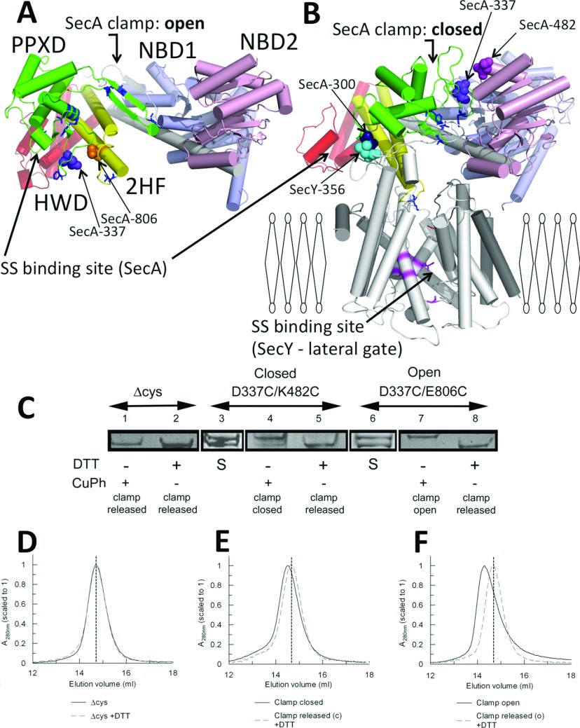
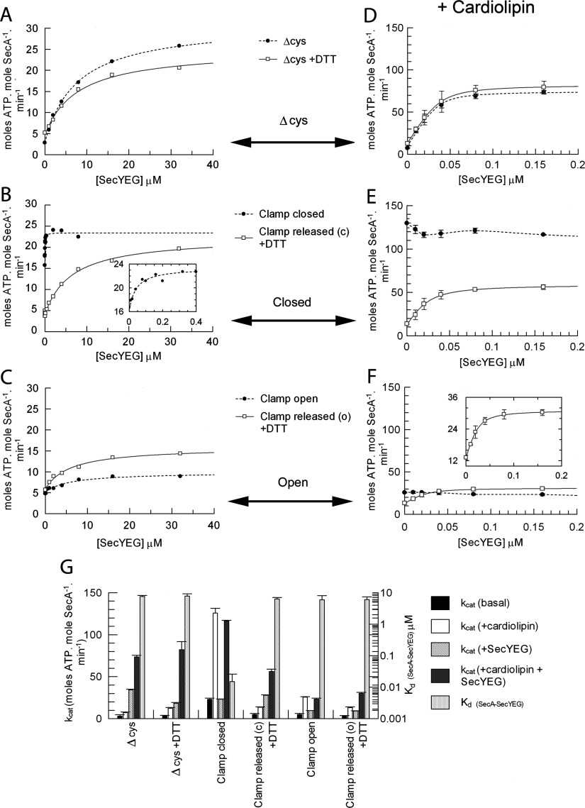
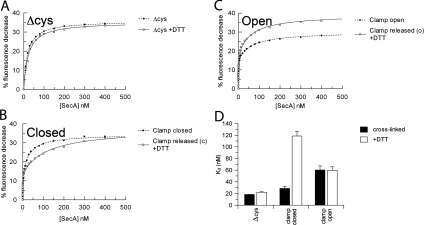
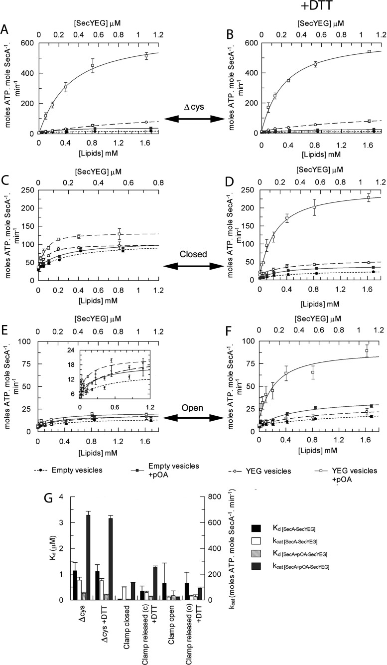
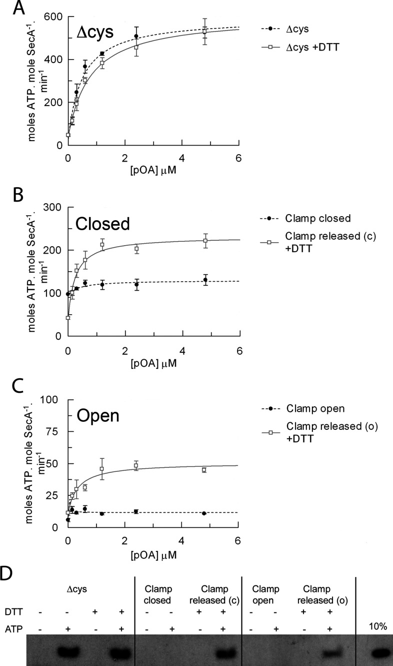
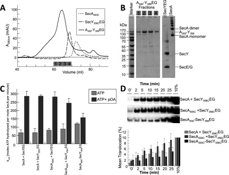
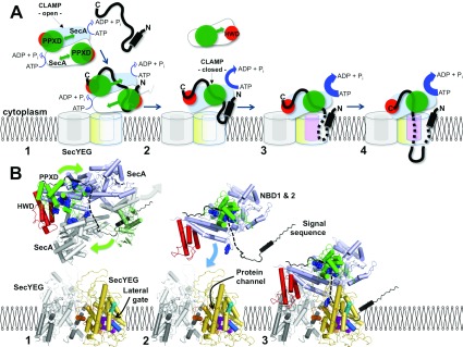
The motor ATPase SecA drives protein secretion through the bacterial Sec complex. The PPXD (pre-protein cross-linking domain) of the enzyme has been observed in different positions, effectively opening and closing a clamp for the polypeptide substrate. We set out to explore the implicated dynamic role of the PPXD in protein translocation by examining the effects of its immobilization, either in the position occupied in SecA alone with the clamp held open or when in complex with SecYEG with the clamp closed. We show that the conformational change from the former to the latter is necessary for high-affinity association with SecYEG and a corresponding activation of ATPase activity, presumably due to the PPXD contacting the NBDs (nucleotide-binding domains). In either state, the immobilization prevents pre-protein transport. However, when the PPXD was attached to an alternative position in the associated SecYEG complex, with the clamp closed, the transport capability was preserved. Therefore large-scale conformational changes of this domain are required for the initiation process, but not for translocation itself. The results allow us to refine a model for protein translocation, in which the mobility of the PPXD facilitates the transfer of pre-protein from SecA to SecYEG.


Figures

Figure 1
Figure 1. Interactions of the PPXD
SecA protomer from a structure determined as (A) a dimer [6] and (B) a monomer bound to SecYEG (white) [7], viewed from the side of the membrane, with the α-helices shown as cylinders. NBD1 is shown in blue, NBD2 in pink, the HSD including 2HF in yellow, the PPXD in green and the HWD in red. The signal-sequence-binding sites are shown for SecA [15] and SecY [20]. Specific residues used for cross-linking are shown in space-fill representation, and labelled according to the E. coli residue numbers: SecAD337C/E806C (clamp open; depicted in A), SecAD337C/K482C (clamp closed; depicted in B), SecA300–SecY356EG (clamp closed; depicted in B). (C) Gradient SDS/PAGE was used to resolve the intramolecular cross-links in the presence of oxidizing (1 mM copper phenanthroline) or reducing (10 mM DTT) agent. Lanes 1 and 2, SecAΔcys; lanes 3–5, SecAD337C/K482C; lanes 6–8, SecAD337C/E806C. S denotes the starting material, which contained both species [cross-linked (top band) and uncross-linked (bottom band)]. (DF) The corresponding size-exclusion chromatography analysis for each sample is shown below: (D) SecAΔcys±DTT; (E and F) cross-linked and uncross-linked mutants (black continuous traces or grey broken traces respectively) used to trap the clamp in the closed and open states. A vertical broken line shows the elution volume of SecAΔcys in all three traces. CuPh, copper phenanthroline.
Figure 2
Figure 2. Analysis of SecA ATPase activity and binding affinity for SecYEG
Steady-state ATPase activity of 0.15 μM SecA in TKM buffer in the presence of 1 mM ATP and increasing concentrations of purified detergent-solubilized SecYEG. Data were fitted to a ligand-binding equation and the parameters are shown in Supplementary Table S1 (at http://www.BiochemJ.org/bj/449/bj4490695add.htm), columns 1 and 2. (A) SecAΔcys in the absence (broken trace) and presence (continuous trace) of 10 mM DTT. (B) SecAD337C/K482C cross-linked with the clamp closed (broken trace; no DTT) and with the clamp released from the closed (c) state (continuous trace; 10 mM DTT). (C) SecAD337C/E806C cross-linked with the clamp open (broken trace; no DTT) and with the clamp released from the open (o) state (continuous trace; 10 mM DTT). (DF) Experiments shown in (AC) were repeated in the presence of 40 μM CL. (G) The end point kcat values under the conditions described, along with the Kd of SecA binding SecYEG, are shown in the histogram (also in Supplementary Table S1, columns 3 and 4).
Figure 3
Figure 3. Measurement of the binding of the SecA 2HF to SecY268FlEG
SecA was titrated into a solution containing 5 nM SecY268FlEG and the fluorescence quench expressed as the percentage fluorescence decrease. (A) SecAΔcys in the absence (broken trace) and presence (continuous trace) of 10 mM DTT. (B) SecAD337C/K482C in the absence (broken trace; clamp closed) and presence [continuous trace; clamp released from closed (c)] of 10 mM DTT. (C) SecAD337C/E806C in the absence (broken trace; clamp open) and presence [continuous trace; clamp released from open (o)] of DTT. (D) Parameters from the fits are shown in the histogram (also in Supplementary Table S1 at http://www.BiochemJ.org/bj/449/bj4490695add.htm, columns 7 and 8).
Figure 4
Figure 4. Analysis of the effect of proOmpA on the binding affinity between SecA and SecYEG
The steady-state ATPase activity of 80 nM SecA in TKM buffer was measured in the presence of 1 mM ATP and increasing amounts of either empty vesicles (● and , bottom axis) or vesicles reconstituted with purified SecYEG (○ and □, top axis) in the absence or presence of 0.6 μM proOmpAΔcys (circles compared with squares respectively). (A) SecAΔcys, (B) SecAΔcys+10 mM DTT, (C) SecAD337C/K482C with the clamp locked closed, (D) SecAD337C/K482C with the clamp released from closed (c)+DTT, (E) SecAD337C/E806C with the clamp locked open, (F) SecAD337C/E806C with the clamp released from the open state (o)+DTT. (G) Data were fitted to a ligand-binding equation and the parameters are shown in the histogram (also in Supplementary Table S1 at http://www.BiochemJ.org/bj/449/bj4490695add.htm, columns 9–12).
Figure 5
Figure 5. Intramolecular immobilization of the PPXD within SecA inhibits protein translocation
Steady-state ATPase activity of 80 nM SecA in TKM buffer in the presence of 1 mM ATP and 1.08 μM SecYEG reconstituted into E. coli polar lipids, and increasing amounts of purified proOmpAΔcys. (A) SecAΔcys in the absence (broken trace) and presence (continuous trace) of 10 mM DTT. (B) SecAD337C/K482C in the absence (broken trace; clamp closed) and presence [continuous trace; clamp released from the closed state (c)] of 10 mM DTT. (C) SecAD337C/E806C in the absence (broken trace; clamp open) and presence [continuous trace; clamp released from the open state (o)] of DTT. Data were fitted to a ligand-binding equation and the parameters are shown in Supplementary Table S1 at http://www.BiochemJ.org/bj/449/bj4490695add.htm, columns 13 and 14. (D) In vitro translocation reactions were carried out from end point ATPase assays. Successfully translocated proOmpA was detected via immunoblot analysis.
Figure 6
Figure 6. Purification and analysis of the cross-linked SecA–SecYEG complex
(A) Size-exclusion chromatography: SecY356CEG, SecA300C and SecA300–Y356EG. Fractions analysed by SDS/PAGE in (B) are indicated by the numbered grey boxes. (B) SDS/PAGE of the purified cross-linked SecA300–Y356EG complex contained in adjacent fractions eluting from the final size-exclusion purification step. SecYEG and a SecA sample containing monomers and SDS-resistant dimers (and other higher aggregated states) were run as additional gel markers (not as representatives of the input to the cross-linking experiment). (C) Steady-state ATPase activity of 0.3 μM wild-type SecA or SecA300C with saturating (1 μM) wild-type SecYEG or SecY356CEG proteoliposomes, or the cross-linked complex SecA300–Y356EG reconstituted into phospholipid vesicles, with and without 0.7 μM proOmpA. The results were averaged from four independent experiments. (D) Top panel: relative levels of translocation for wild-type SecA or SecA300C with SecY356CEG proteoliposomes, or the cross-linked complex (SecA300–Y356EG) proteoliposomes over 25 min, analysed by anti-proOmpA immunoblot. t=‘0’ represents translocation achieved after initial mixing of reaction components, approximately 10 s. The blot is representative of n=5, which were quantified using ImageJ (NIH) software (bottom panel).
Figure 7
Figure 7. Model depicting the role of the PPXD in protein translocation
(A) Schematic diagram and (B) structural (cartoon) representation of various states of the translocation cycle of SecA and SecYEG. SecYEG: non-translocation complex (grey), translocating complex (yellow), within (B) TMS2b (cyan cylinder) and TMS7 (blue cylinder) of the lateral gate, as well as the residues that contact substrate (magenta space-fill) [20]. SecA: two monomers of the dimer (pale blue and white), PPXD (green), HWD (red) and in (B) residues that contact substrate (blue space-fill) [15]. Pre-protein (black) with the N-terminal signal sequence (black cylinder). Stage (1) initiation: SecA dimers (low ATPase activity [23]) engage SecYEG. The mobility of the PPXD permits the binding of the pre-protein between the PPXD and the HWD. In (B) the structure of the SecA dimer is shown [6], where the lower monomer has been replaced by SecA (white with PPXD in pale green) bound to the signal sequence (black) [14]. In the upper monomer the C-terminus is shown in black, which may occupy the pre-protein-binding site [27]. The broken black line connects this C-terminal stretch to the signal sequence, describing a possible continuous binding groove for the pre-protein. SecYEG here and in subsequent stages (2) and (3) was modelled on the structure of the membrane-bound complex [4,9]. Stage (2) activation: dissociation of SecA [18] exposes the SecYEG-binding site of SecA [7]. The relocation of the PPXD serves to increase the affinity for SecYEG, activate the ATPase activity [19], release the signal sequence from the departing SecA and close the clamp about the pre-protein. In (B) the dissociated SecA is removed but the signal sequence is retained. The remaining monomeric enzyme has been replaced by the activated version seen in the SecA–SecYEG complex [7], with the PPXD adjacent to the NBDs to trap the pre-protein. Stage (3) insertion: the resultant association of the monomeric SecA with SecYEG displays the signal sequence to the binding site at the lateral gate of SecY [8,28]. The binding of which unlocks SecYEG [8] to promote the intercalation of the translocation substrate into the protein channel and the full activation of the ATPase [19]. In (B) the model of the membrane-bound translocon is shown [7,9] retaining the pre-protein from the previous alignment. Stage (4) transport: the trapped and inserted pre-protein passes through the membrane via a single SecYEG complex.

References

    1. Blobel G., Dobberstein B. Transfer of proteins across membranes. I. Presence of proteolytically processed and unprocessed nascent immunoglobulin light chains on membrane-bound ribosomes of murine myeloma. J. Cell Biol. 1975;67:835–851. - PMC - PubMed
    1. Gouridis G., Karamanou S., Gelis I., Kalodimos C. G., Economou A. Signal peptides are allosteric activators of the protein translocase. Nature. 2009;462:363–367. - PMC - PubMed
    1. Brundage L., Hendrick J. P., Schiebel E., Driessen A. J., Wickner W. The purified E. coli integral membrane protein SecY/E is sufficient for reconstitution of SecA-dependent precursor protein translocation. Cell. 1990;62:649–657. - PubMed
    1. Breyton C., Haase W., Rapoport T. A., Kuhlbrandt W., Collinson I. Three-dimensional structure of the bacterial protein-translocation complex SecYEG. Nature. 2002;418:662–665. - PubMed
    1. van den Berg B., Clemons W. M., Jr, Collinson I., Modis Y., Hartmann E., Harrison S. C., Rapoport T. A. X-ray structure of a protein-conducting channel. Nature. 2004;427:36–44. - PubMed

Publication types

MeSH terms

LinkOut - more resources