. 2012 Nov 5:13:140.
doi: 10.1186/1471-2202-13-140.

Visualization of odor-induced neuronal activity by immediate early gene expression

Affiliations

Visualization of odor-induced neuronal activity by immediate early gene expression

Asim K Bepari et al. BMC Neurosci. .

Abstract

Background: Sensitive detection of sensory-evoked neuronal activation is a key to mechanistic understanding of brain functions. Since immediate early genes (IEGs) are readily induced in the brain by environmental changes, tracing IEG expression provides a convenient tool to identify brain activity. In this study we used in situ hybridization to detect odor-evoked induction of ten IEGs in the mouse olfactory system. We then analyzed IEG induction in the cyclic nucleotide-gated channel subunit A2 (Cnga2)-null mice to visualize residual neuronal activity following odorant exposure since CNGA2 is a key component of the olfactory signal transduction pathway in the main olfactory system.

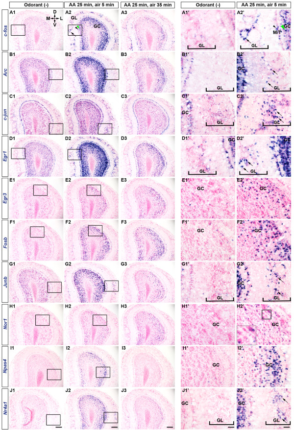
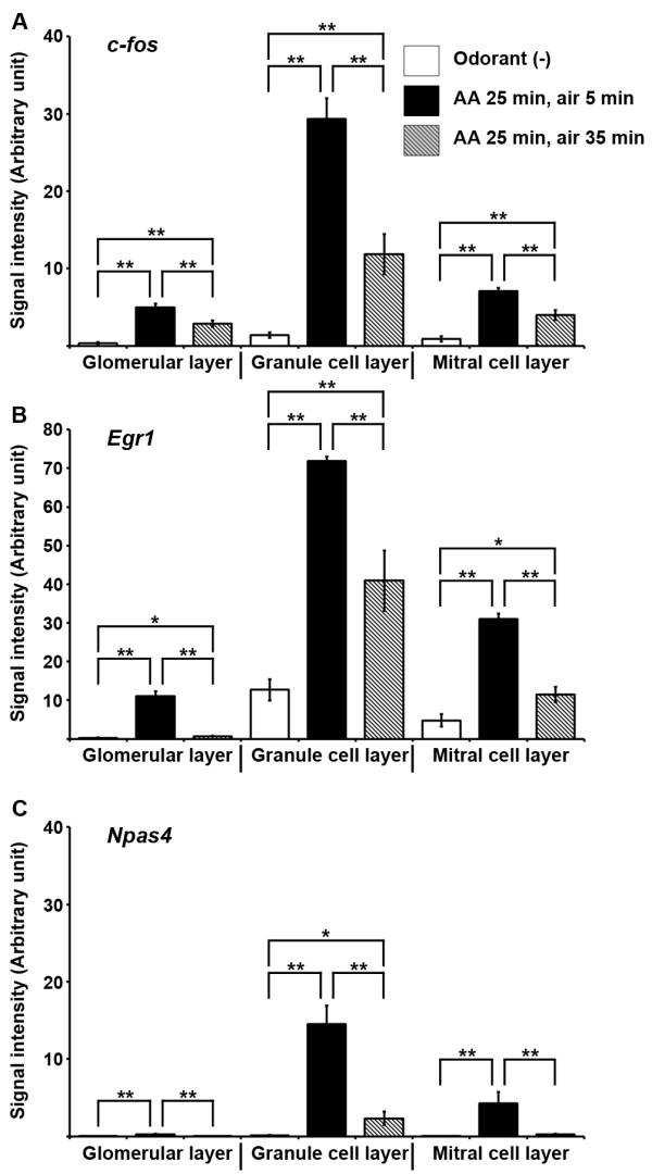
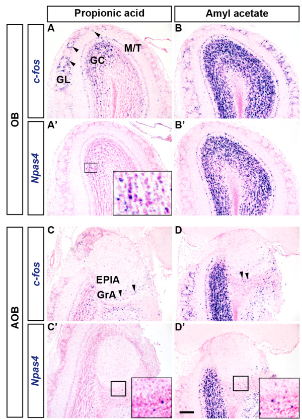
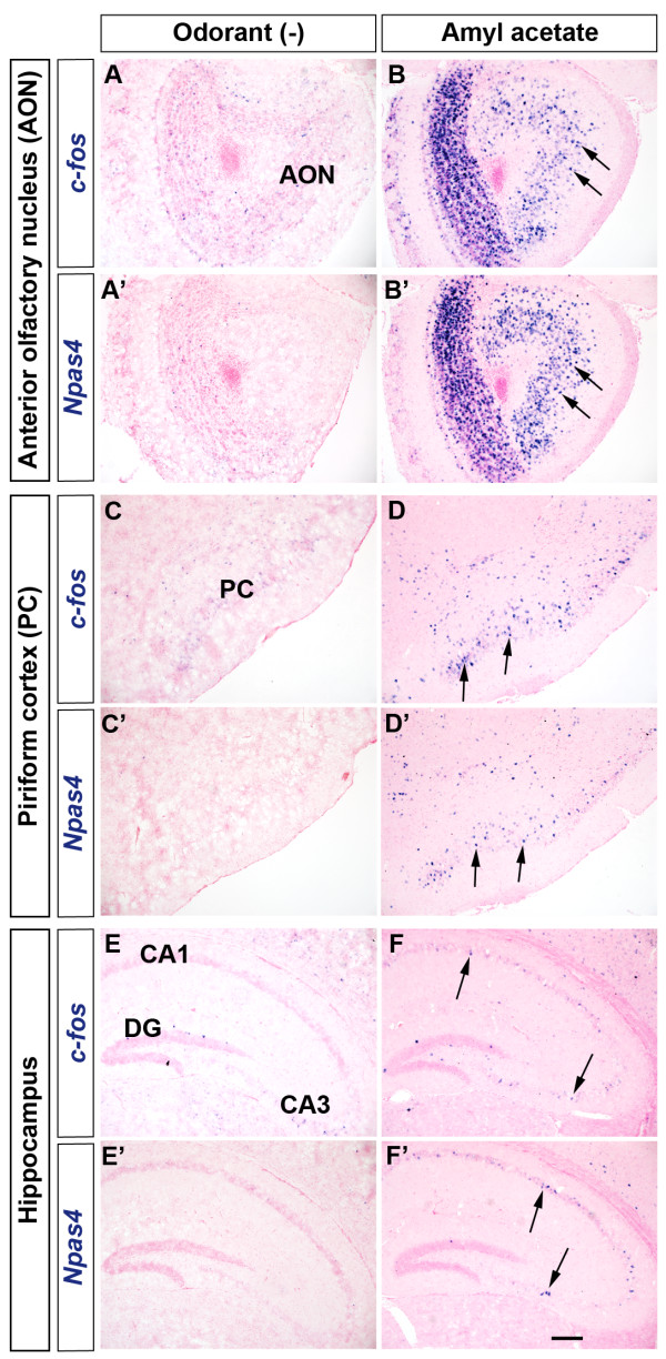
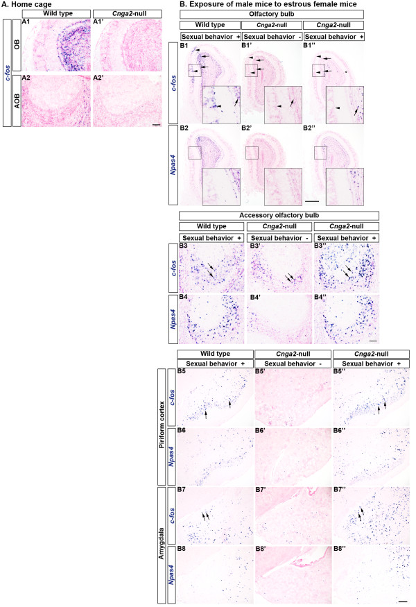
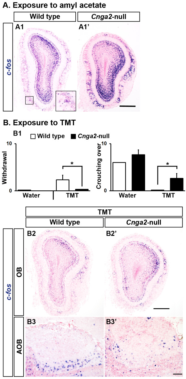
Results: We observed rapid induction of as many as ten IEGs in the mouse olfactory bulb (OB) after olfactory stimulation by a non-biological odorant amyl acetate. A robust increase in expression of several IEGs like c-fos and Egr1 was evident in the glomerular layer, the mitral/tufted cell layer and the granule cell layer. Additionally, the neuronal IEG Npas4 showed steep induction from a very low basal expression level predominantly in the granule cell layer. In Cnga2-null mice, which are usually anosmic and sexually unresponsive, glomerular activation was insignificant in response to either ambient odorants or female stimuli. However, a subtle induction of c-fos took place in the OB of a few Cnga2-mutants which exhibited sexual arousal. Interestingly, very strong glomerular activation was observed in the OB of Cnga2-null male mice after stimulation with either the neutral odor amyl acetate or the predator odor 2, 3, 5-trimethyl-3-thiazoline (TMT).

Conclusions: This study shows for the first time that in vivo olfactory stimulation can robustly induce the neuronal IEG Npas4 in the mouse OB and confirms the odor-evoked induction of a number of IEGs. As shown in previous studies, our results indicate that a CNGA2-independent signaling pathway(s) may activate the olfactory circuit in Cnga2-null mice and that neuronal activation which correlates to behavioral difference in individual mice is detectable by in situ hybridization of IEGs. Thus, the in situ hybridization probe set we established for IEG tracing can be very useful to visualize neuronal activity at the cellular level.


Figures

Figure 1
Figure 1
Odorant (amyl acetate) exposure induced the expression of IEGs in the mouse OB. Mice were exposed to overhead airflow for 2 h and then to the test odorant (amyl acetate) for 25 min (5-min exposures with 5-min intervals). The ISH of coronal sections of OB indicated low expression levels of ten IEGs in mice immediately after the 2-h air exposure, (Odorant (−), A1-J1, A1’-J1’). All these ten IEGs were induced in the mouse OB after 30 min of odor onset (AA 25 min, air 5 min, A2-J2, A2’-J2’). Boxed areas in A1-J1 and A2-J2 are magnified in A1’-J1’ and A2’-J2’, respectively. Inset in H2’ is a magnified view of the boxed area. Odor-evoked induction of IEG expression was transient and expression levels of most of the IEGs declined within 60 min of initial odorant exposure (AA 25 min, air 35 min, A3-J3). Arrows indicate GL, black arrowheads indicate M/T and green arrowheads indicate GC. AA, amyl acetate; GL, Glomerular layer; M/T, Mitral/Tufted cell layer; GC, Granule cell layer. D, Dorsal; V, Ventral; M, Medial; L, Lateral. Scale bars: (A1-J3) 200 μm, (A1’-J2’) 50 μm.
Figure 2
Figure 2
Quantification of odor-evoked IEG induction in the mouse OB. Signal intensity (arbitrary unit) of c-fos (A), Egr1 (B) and Npas4 (C) was calculated as the percentage of area positive for ISH signals in respective layers of the OB. Columns represented mean ± SEM. Seven to eight bulbs (approximately from + 4.5 mm bregma to + 4 mm bregma) from two to three mice were analyzed. Student's t-test was performed to compare means. ** Difference between groups was highly significant (p ≤ 0.01). * Difference between groups was significant (p ≤ 0.05).
Figure 3
Figure 3
Comparison of IEG induction patterns in response to two different odorants. Mice were sacrificed after the 30-min continuous exposure to the test odorant. (A, A’) Propionic acid activated several glomeruli specifically in the dorsal OB (arrowheads in A). Induced expression of Npas4 was observed only in the granule cell layer (A’, inset). (B, B’) A large number of glomeruli were activated by amyl acetate. Npas4 induction was apparent only in the granule cell layer (B’). (C-D’) Patterns of IEG induction in the AOB after odorant exposure. Arrowheads indicate c-fos induction in the granule cell layer of the AOB (C, D). Only a slight induction of Npas4 was observed in the AOB (C’, D’, insets). GL, Glomerular layer; M/T,Mitral/Tufted cell layer; GC, Granule cell layer; GrA, Granule cell layer of the AOB; EPlA, External plexiform layer of the AOB. Scale bar: 200 μm.
Figure 4
Figure 4
Odorant exposure induced activity-dependent gene expression in different brain regions. Odorant exposure induced expression of c-fos (A-F) and Npas4 (A’-F’) in the AON (arrows, A-B’), the PC (arrows, C-D’) and the hippocampus (arrows, E-F'). DG, Dentate gyrus. Scale bar: 200 μm.
Figure 5
Figure 5
Individual differences in induction of activity-dependent genes in Cnga2-null mice after exposure to female mice.A. Expression of c-fos in mice which were sacrificed from their home cages without any odorant exposure. Significantly reduced expression levels of c-fos were observed in the OB (A1’) and AOB (A2’) of Cnga2-null male mice compared to that of wild type male littermates (A1, A2, respectively). B. Induction of c-fos expression in male mice which were exposed to estrous female mice. Arrowheads indicate the glomerular layer and arrows indicate the granule cell layer. Sexual stimulation by female mice induced expression of IEGs in the wild type OB (B1, B2). IEG induction was almost absent in the Cnga2 mutants which did not show sexual behaviors (B1’, B2’). IEG induction occurred in the OB, mainly in the granule cell layer, of the Cnga2-null male mice which showed sniffing and mounting behaviors (B1”, B2”). Insets in (B1-B2”) show magnified views of the boxed areas. IEG induction occurred in the AOB of male mice exposed to female mice (arrows, B3-B4”). (B5-B6”) Induction of IEGs in the PC (arrows) after exposure to female mice. Both in the wild type mice (B5, B6) and the mutants (B5”, B6”) which showed sexual behaviors, expression of IEGs was induced in the PC. IEG induction did not occur in the PC of Cnga2-null mice (B5’, B6’) which did not show sexual behaviors. (B7-B8”) Induction of IEGs in the MePD (arrows) after exposure to female mice. Both in the wild type mice (B7, B8) and the mutants (B7”, B8”) which showed sexual behaviors, expression of IEGs was induced in the MePD. The IEG induction did not occur in the MePD of Cnga2-null mice (B7’, B8’) which did not show sexual behaviors. Scale bars: (A1-A2’ and B3-B4”) 100 μm, (B1-B2”) 500 μm, (B5-B8’) 200 μm.
Figure 6
Figure 6
Neuronal activation in response to amyl acetate and TMT in Cnga2-null mice. A. A neutral odorant, amyl acetate, robustly induced c-fos expression in the OB in both wild type (A1) and Cnga2-null (A1’) mice. Inset in A1 shows magnified view of the boxed area. B. Responses of mice after presentation of TMT, a predator odor from fox. TMT-induced avoidance behaviors were present in wild type mice but absent in Cnga2-null mice (number of withdrawal, B1 left). Unlike wild type mice, Cnga2-null mice showed increased investigating behaviors for TMT (number of crouching over, B1 right). After exposure to TMT for 30 min, expression of c-fos was induced in both wild type and Cnga2-null mice in the OB (B2, B2’, respectively) and the AOB (B3, B3’, respectively). Scale bars: (A1, A1’, B2, B2’) 500 μm, (B3-B3’) 100 μm.

References

    1. Flavell SW, Greenberg ME. Signaling mechanisms linking neuronal activity to gene expression and plasticity of the nervous system. Annu Rev Neurosci. 2008;31:563–590. doi: 10.1146/annurev.neuro.31.060407.125631. - DOI - PMC - PubMed
    1. Norlin EM, Vedin V, Bohm S, Berghard A. Odorant-dependent, spatially restricted induction of c-fos in the olfactory epithelium of the mouse. J Neurochem. 2005;93:1594–1602. doi: 10.1111/j.1471-4159.2005.03159.x. - DOI - PubMed
    1. Busto GU, Elie JE, Kermen F, Garcia S, Sacquet J, Jourdan F, Marcel D, Mandairon N, Didier A. Expression of Zif268 in the granule cell layer of the adult mouse olfactory bulb is modulated by experience. Eur J Neurosci. 2009;29:1431–1439. doi: 10.1111/j.1460-9568.2009.06689.x. - DOI - PubMed
    1. Okuno H. Regulation and function of immediate-early genes in the brain: beyond neuronal activity markers. Neurosci Res. 2011;69:175–186. doi: 10.1016/j.neures.2010.12.007. - DOI - PubMed
    1. Wisden W, Errington ML, Williams S. Differential expression of immediate early genes in the hippocampus and spinal cord. Neuron. 1990;4:603–614. doi: 10.1016/0896-6273(90)90118-Y. - DOI - PubMed

Publication types

MeSH terms

Substances