

Multiscale forward electromagnetic model of uterine contractions during pregnancy
- PMID: 23126570
- PMCID: PMC3605117
- DOI: 10.1186/1756-6649-12-4
Multiscale forward electromagnetic model of uterine contractions during pregnancy
Abstract
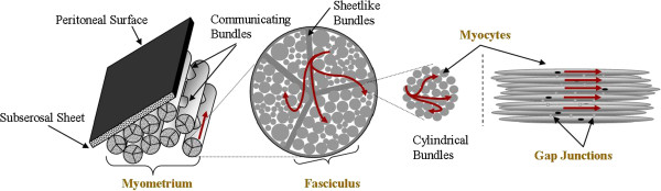
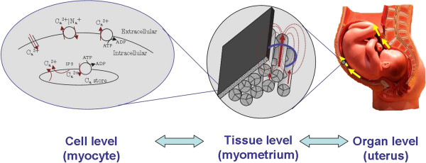
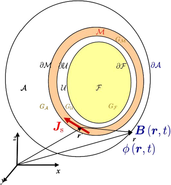
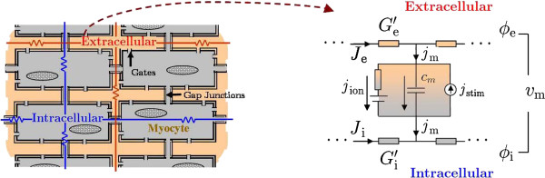
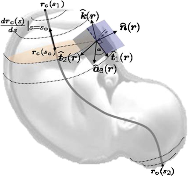
Background: Analyzing and monitoring uterine contractions during pregnancy is relevant to the field of reproductive health assessment. Its clinical importance is grounded in the need to reliably predict the onset of labor at term and pre-term. Preterm births can cause health problems or even be fatal for the fetus. Currently, there are no objective methods for consistently predicting the onset of labor based on sensing of the mechanical or electrophysiological aspects of uterine contractions. Therefore, modeling uterine contractions could help to better interpret such measurements and to develop more accurate methods for predicting labor. In this work, we develop a multiscale forward electromagnetic model of myometrial contractions during pregnancy. In particular, we introduce a model of myometrial current source densities and compute its magnetic field and action potential at the abdominal surface, using Maxwell's equations and a four-compartment volume conductor geometry. To model the current source density at the myometrium we use a bidomain approach. We consider a modified version of the Fitzhugh-Nagumo (FHN) equation for modeling ionic currents in each myocyte, assuming a plateau-type transmembrane potential, and we incorporate the anisotropic nature of the uterus by designing conductivity-tensor fields.
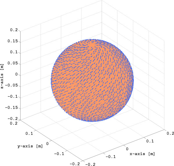
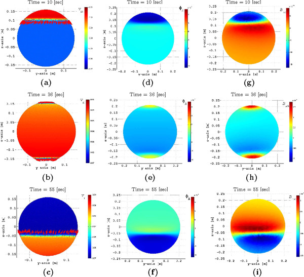
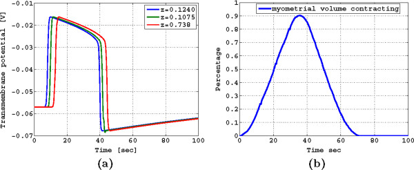
Results: We illustrate our modeling approach considering a spherical uterus and one pacemaker located in the fundus. We obtained a travelling transmembrane potential depolarizing from -56 mV to -16 mV and an average potential in the plateau area of -25 mV with a duration, before hyperpolarization, of 35 s, which is a good approximation with respect to the average recorded transmembrane potentials at term reported in the technical literature. Similarly, the percentage of myometrial cells contracting as a function of time had the same symmetric properties and duration as the intrauterine pressure waveforms of a pregnant human myometrium at term.
Conclusions: We introduced a multiscale modeling approach of uterine contractions which allows for incorporating electrophysiological and anatomical knowledge of the myometrium jointly. Our results are in good agreement with the values reported in the experimental technical literature, and these are potentially important as a tool for helping in the characterization of contractions and for predicting labor using magnetomyography (MMG) and electromyography (EMG).
Figures

References
-
- Chard T, Grudzinskas JG. The Uterus. Cambridge: University Press; 1995.
-
- Degani S, Leibovitz Z, Shapiro I, Gonen R, Ohel G. Myometrial thickness in pregnancy: longitudinal sonographic study. J Ultrasound Med. 1998;17(10):661–665. - PubMed
-
- Weiss S, Jaermann T, Schmid P, Staempli P, Niederer P, Caduff R, Bajka M. Three-dimensional fiber architecture of the nonpregnant human uterus determined Ex Vivo using magnetic resonance diffusion tensor imaging. Anat Rec Part A, Discov Mol, Cell, Evol Biol. 2006;288:84–90. - PubMed
LinkOut - more resources
Full Text Sources
